Last Updated on November 22, 2023
Metatarsal Bones or Metatarsus
There are five metatarsal bones in the foot which are numbered from medial to lateral side.
Common Features of Metatarsal Bones

Each metatarsal bone is a miniature long bone and has following parts
Base
Base is the proximal end of metatarsal bone and articulates with tarsal bones. It is set obliquely in such a way that it projects backwards and laterally.
Shaft
The shaft is slightly convex dorsally and concave ventrally [The upper surface of the foot is dorsal and plantar surface or lower surface is ventral.]
Shaft is prismoid in from and tapers from base to the head. The shafts of metatarsal bones give origin to interossei.
Head
Head is the distal end of the metatarsal or distal end. It is flattened from side to side. Head of each metatarsal articulates with respective phalanx to form metatarsophalangeal joint.
A constriction proximal to head is called anatomical neck. A second constriction more proximal on the shaft is known as surgical neck.
The First Metatarsal Bone
First metatarsal is the shortest, thickest and strongest of all metatarsal bones. It is adapted so for transmission of the body weight.
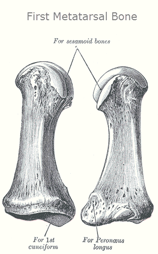
The proximal surface of the base has kidney shaped facet, which is concave outwards. The first metatarsal articulates with the medial cuneiform, and to a small extent to the intermediate cuneiform bone and on distal side with proximal respective phalanx.
The circumference of the kidney shaped facet is grooved to receive tarsometatarsal ligaments.
The base medially provides attachment to part of tibialis anterior tendon.
Generally, there is no articular facet on the sides but sometimes an oval facet might be present to articulate with second metatarsal bone.
A rough oval prominence on the plantar surface is for insertion of peroneus longus, also called fibularis longus.
The body of the bone is strong, and of well-marked prismoid form.
The head is large and has two gooved facets on its plantar surface, separated by an elevation, for articulation with sesamoid bones.
On its plantar surface are two grooved facets, on which glide sesamoid bones, the facets are separated by a smooth elevation.
Second Metatarsal Bone
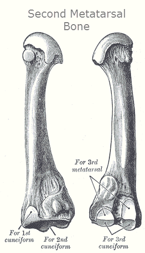
Second metatarsal is the longest metatarsal. It has wedge-shaped base. It projects backwards and is held in the recess formed by the three cuneiform bones.
The medial side of the base has a dorsal facet for the medial cuneiform.
It articulates with the cuneiform bones, third metatarsal and occasionally the first metatarsal bone.
Its base is broad above, narrow and rough below.
It presents four articular surfaces:
- Posterior aspect of the base, a triangular facet is for articulation with the intermediate cuneiform bone
- A facet on upper part of medial surface articulates with medial cuneiform
- On lateral aspect of base, an upper and lower facets are present, separated by a rough non-articular interval. A vertical ridge divides both of these lateral articular surfaces into two. The anterior ones articulate with the third metatarsal and the posterior ones with the lateral cuneiform.
- A fifth facet is occasionally present for articulation with the first metatarsal; it is oval in shape, and is situated on the medial side of the body near the base.
The second metatarsal base acts as a keystone for the Lisfranc joint or tarsometatarsal articulations. A keystone is the wedge-shaped stone piece at the apex of a masonry vault or arch, which is the final piece placed during construction and locks all the stones into position, allowing the arch to bear weight. See the image below to understand keystone concept.
 Third Metatarsal Bone
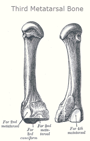
The third metatarsal bone is a long bone in the foot. It is the second longest metatarsal.
The base is wedge shaped.
Articulations
- Third metatarsal bone articulates proximally, by means of a triangular smooth surface, with the third cuneiform
- medially, by two facets, with the second metatarsal
- Laterally a single facet for the fourth metatarsal. This last facet is situated at the dorsal angle of the base.
The head or articulates with the third proximal phalanx.
Fourth Metatarsal Bone

The fourth metatarsal bone is smaller in size than the third metatarsal bone and is the third longest metatarsal.
Base of fourth metatarsal is wedge shaped and has a quadrangular surface to articulate with cuboid.
On lateral side, the base has one dorsal facet for fifth metatarsal bone.
The medial side of the base has one faced placed dorsally, which is subdivided into a proximal part for the lateral cuneiform and a distal part for the third metatarsal bone.
The head articulates with the fourth proximal phalanx.
Fifth Metatarsal Bone
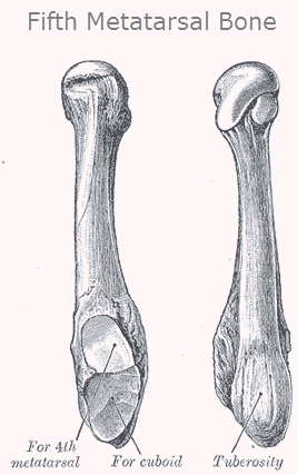
The fifth metatarsal bone is a long bone in the foot. It is the second smallest of the five metatarsal bones. Fifth metatarsal has a large tuberosity or styloid process which projects backwards and laterally.
The base articulates behind, by a triangular surface cut obliquely in a transverse direction.
The medial side of the base has one facet for the fourth metatarsal bone.
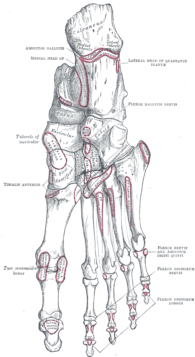
The plantar surface of the base is groove by the tendon of the abductor digit minimi.
The head articulates with the fifth proximal phalanx, the first bone in the fifth toe.
A strong band of the plantar aponeurosis connects the projecting part of the tuberosity with the lateral process of the tuberosity of the calcaneus.
Attachments to Metatarsal Bones
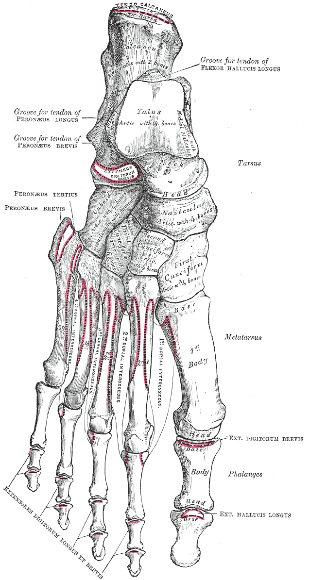
First Metatarsal Bone
- A part of the tibialis anterior is inserted on the medial side of the base of the first metatarsal bones.
- The greater part of the peroneus longus is inserted on a large impression at the inferior angle of the lateral surface of the base of the first metatarsal bone.
- The lateral part of the first dorsal interossei muscle originates from the medial side of the bone. The function of the muscle is to spread the toes
Second Metatarsal Bone
- First and second dorsal interossei muscles attach to the second metatarsal bone.
- The first dorsal interossei from the medial side of the bone and the second dorsal interossei from the lateral side. Dorsal interossei function to spread toes.
- The horizontal head of the adductor hallucis originates from the lateral side of the metatarsophalangeal joint and from the deep transverse metatarsal ligament.
Third Metatarsal Bone
- The second and third dorsal interossei muscles originates from the third metatarsal bone. The second from the medial side and the third from the lateral side.
- The first Plantar interossei muscle originates from the medial side of the base and shaft of the third metatarsal. The function of the muscle is to move the third toe medially and move the toes together.
- The horizontal head of the adductor hallucis also originates from the lateral side of the metacarpophalangeal joint.
Fourth Metatarsal Bone
- The third and fourth dorsal interossei muscles originates from fourth metatarsal bone. The third dorsal interossei from the medial side and the fourth dorsal interossei from the lateral side.
- The second Plantar interossei muscle originates from the medial side of the base and shaft of the fourth metatarsal.
- The horizontal head of the adductor hallucis also originates from the lateral side of the metacarpophalangeal joint.
Fifth Metatarsal Bone
- The peroneus brevis is inserted on the dorsal surface of the tuberosity of the fifth metatarsal bone.
- The peroneus tertius is inserted on the medial part of the dorsal surface of the base and the medial border of the shaft of the fifth metatarsal bone.
- The flexor digiti minimi brevis arises from the plantar surface of the base of the fifth metatarsal bone.
- The plantar surface of the base is grooved for the tendon of the abductor digiti quinti, and gives origin to the flexor digiti minimi brevis.
- The fourth dorsal interossei muscles originates from the medial side of shaft. The function of the muscle is to spread the toes.
- The third Plantar interossei muscle originates from the medial side of the base and shaft of the fifth metatarsal. The function of the muscle is to move the fourth toe medially and move the toes together.
- The horizontal head of the adductor hallucis from the deep transverse metatarsal ligament
Ossification of Metatarsal Bones
Each metatarsal bone ossifies from one primary and one secondary center.
- Primary centre – Appears in the shaft during the 10th week of the fetal life in the first metatarsal, and during the 9th week fetal life in the rest of the metatarsal.
- Secondary centers
o Base of the first metatarsal – 3rd year
o Other Metatarsal 3-4 years
o A separate center for the tuberosity of the fifth metatarsal bone may be present.
o Secondary centers unite with the shaft by 17-20 years.
Anatomy of Phalanges
There are 14 phalanges in each foot; 2 for the great toe and three for each of the other toes. As compared to the phalanges of the hand, these are much smaller in size, and the shafts ( particularly of otherwise their arrangement and features are similar in the two limbs.
Attachments on Phalanges
Bases of distal phalanges
- Lateral four toes
- Flexor digitorum longus on the plantar surface
- Exetensor expansion on the dorsal surface.
- Great toe
- Flexor Hallucis longus on the plantar surface
- Extensor hallucis longus on the dorsal surface.
Bases of Middle Phalanges
- Flexor digitorum brevis on plantar surface
- Extensor expansion on the dorsal surface.
Bases of Proximal Phalanges
- 2nd, 3rd and 4th toes:
- Lumbrical muscle on the medial side
- Interosseus muscle on each side.
- Fifth Toe
- Plantar interosseus muscle on the medial side
- Abuductor digit minimi and the flexor digit minimi brevis on the lateral side.
- Great toe
- Abductor hallucis and a part of the flexor hallucis medially
- Abductor hallucis and the remaining part of the flexor hallucis brevis laterally.
Fibrous flexor sheath is attached to the margins of the proximal and middle phalanges of the lateral four toes.
Sesamoid Bones in the Foot
- There is one sesamoid bone in the tendon of the peroneus longus. It articulates with the cuboid.
- There are two small sesamoids in the tendons of the flexor hallucis brevis. They articulate with the head of the first metatarsal in the bone.
- Sesamoid bones may be present in the tendons of
- Tibialis anterior
- Tibialis posterior
- Tendons crossing the metatasro-phalangeal and interphalangeal joints
Articulations of Metatarsal Bones and Phalanges
Tarsometatarsal articulations or Lisfranc joint are arthrodial joints in the foot. The tarsometatarsal articulations involves the first, second and third cuneiform bone, the cuboid bone and the metatarsal bones.
- First metatarsal bone articulates with the first cuneiform
- Second metatarsal bone is deeply wedged in between the first and third cuneiforms articulating by its base with the second cuneiform
- Third metatarsal articulates with the third cuneiform
- Fourth metatarsal articulates with the cuboid and third cuneiform
- Fifth metatarsal bone articulate with the cuboid.
Ligaments
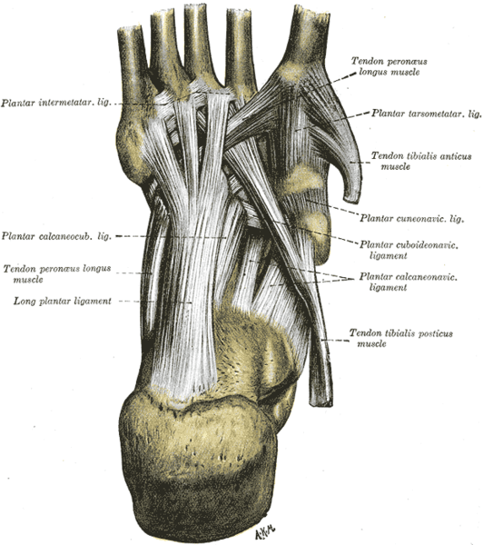
The bones are connected by dorsal, plantar, and interosseous ligaments.

The Dorsal Ligaments
The dorsal ligaments are strong, flat bands.
- The first metatarsal is joined to the first cuneiform by a broad, thin band
- Second metatarsal has three bands, one from each cuneiform bone
- Third bone has one band from third cuneiform
- Fourth metatarsal has one from the third cuneiform and one from the cuboid
- Fifth metatarsal has one band from the cuboid.
The Plantar Ligaments
The plantar ligaments consist of longitudinal and oblique bands.
- First and second metatarsals are the strongest ligaments.
- Second and third metatarsals are joined by oblique bands to the first cuneiform
- Fourth and fifth metatarsals are connected by a few fibers to the cuboid.
The Interosseous Ligaments
The interosseous ligaments are three in number.
- The first interosseous ligament is the strongest. It passes from the lateral surface of the first cuneiform to the adjacent angle of the second metatarsal.
- Second interosseous ligament connects the third cuneiform with the adjacent angle of the second metatarsal.
- Third interosseous ligament is from lateral angle of the third cuneiform to the adjacent side of the base of the third metatarsal.
Movements
Only limited to slight gliding of the bones upon each other is permitted.
Metatarsophalangeal Articulations
The metatarsophalangeal articulations are the joints between the metatarsal bones of the foot and the proximal phalanges of the toes. They are condyloid joints. An elliptical or rounded surface of the metatarsal bones come close to the shallow cavities of the proximal phalanges.
The ligaments are the plantar and two collateral.
Movements
Following movements occur in metatarsophalangeal joints
- Flexion
- Extension
- Abduction
- Adduction
- Circumduction
Clinical Significance
The second metatarsal bone elongation or Morton’s Toe leads to extra-inversion of the foot and putting more stress on knee and back.
Metatarsal bones and phalanges are prone to be fractured.
Injury to tarsometatarsal joints or Lisfranc injury is a serious injury often requiring surgical intervention.
Jones fracture is fracture of base of fifth metatarsal and is a common fracture
Beckham Bone
The Beckham bone is a name attributed by British journalists to the second metatarsal.David Beckham, while playing for Manchester United broke the second metatarsal and for some time the bone in popular media was called Beckham Bone.