• Skip to main content
  • Skip to primary sidebar
  • Skip to footer
  • General Ortho
  • Procedures
  • Spine
  • Upper Limb
  • Lower Limb
  • Pain
  • Trauma
  • Tumors
  • Newsletter/Updates
  • About Us
  • Contact Us

Bone and Spine

Orthopedic health, conditions and treatment

Anatomy of Patella

By Dr Arun Pal Singh

In this article
    • Structure
    • Attachments
    • Blood supply 
    • Determination of side of Patella
    • Clinical Significance of Patella
      • Lateral dislocation of the patella

The patella or kneecap is the sesamoid that lies within the quadriceps tendon/patellar ligament and forms part of the knee joint and is situated in front of the lower end of femur appx 1 cm above the knee joint. If it lies higher, it is called patella alta and if it is lower, it is called patella baja. It is the largest sesamoid bone of the body. The ossification centers of the kneecap appear between 3 and 6 years. They fuse at puberty, with higher levels of activity.

knee and patella


Structure

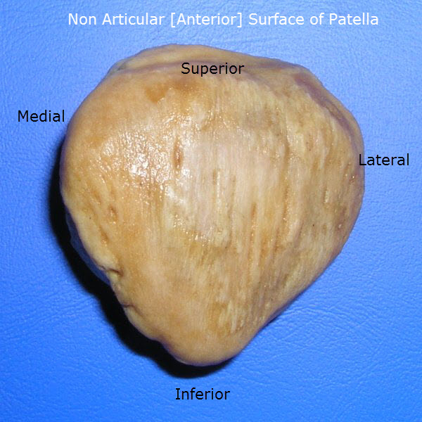
The patella is triangular in shape with a superior base and inferior apex. The apex lies about 1 cm from the knee joint. It has three borders – superior, lateral and medial, and two surfaces – anterior and posterior.

The anterior surface is convex, rough and ridged vertically. It is covered by expansion from tendons of rectus femoris and is separated from the skin by the prepatellar bursa.

Antrior aspect of patella

The posterior surface is smooth, composed of articular cartilage, and is divided into medial and lateral facets. The posterior surface is articular in upper three fourths and nonarticular in its lower one fourth.

Posterior surface of patella

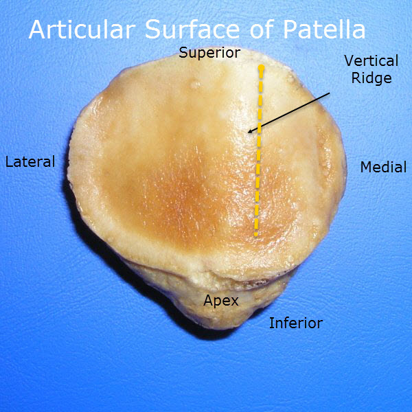
The articular area is divided by a vertical ridge into larger lateral and a smaller medial portion. Another vertical ridge separates a medial strip from the medial portion.

This medial strip articulates with a reciprocal strip on the medial side of intercondylar notch of the femur during full flexion.

The rest of the medial and lateral portions of the articular surface are divided by two transverse lines into three pairs of facets.

The medial and lateral facets of the patella articulate with the medial and lateral condyles of the femur, respectively, to form the patellofemoral component of the knee joint.

Attachments

The base [superior aspect or base of the triangle] provides insertion to rectus femoris in front and to vastus intermedius behind. The lateral border provides insertion to vastus lateralis in its upper one third to half. The medial border provides insertion to vastus lateralis in its upper third to half.

Behind the apex, on the posterior aspect, there is a nonarticular area which provides attachment to the patellar ligament or ligamentum patellae which get attached to the tibial tubercle, inferiorly.

Blood supply 

Arterial blood enters via the anterior surface of the patella and an anastomotic patella ring is formed supplied by the paired superior and inferior geniculate arteries.

Determination of side of Patella

  • A triangular with the apex of the triangle directed downwards.
  • The anterior surface is rough and nonarticular. The upper three-fourths of the posterior surface is smooth and articular.

The bone rests on a broad lateral area when placed on a table as a vertical ridge divides it into the large lateral area and a smaller medial area.

During different phases of movements of the knee, different portions of the patellar bone articulate with the femur. The lower pair of articular facets articulates during extension, middle during flexion and upper pair during mid-flexion. Medial strip articulates during full flexion.

Clinical Significance of Patella

Lateral dislocation of the patella

The quadriceps muscle pulls the patella obliquely and laterally in relation to the femur. There are factors that prevent such displacement: larger lateral condyle of the femur, tension in the medial retinacular fibers and the direction of insertion of fibers of the vastus medialis muscle.

There is a natural tendency in patella to dislocate laterally because of outward angulation of between femur and tibia. This is prevented by the lateral edge of the patellar articular surface being deeper than the medial edge. Moreover, vastus medialis inserts to little lower than vastus lateralis and keep the check.

A disturbance in bony anatomy or muscular balance may result in recurrent dislocation of the patella.

Injury

It is a common bone to be fractured.

Bipartite Patella

It is a condition where there is a failure of fusion of patella.


Spread the Knowledge
  • 3
    Shares
  •  
    3
    Shares
  • 3
  •  
  •  
  •  
  •  

Filed Under: Anatomy

About Dr Arun Pal Singh

Arun Pal Singh is an orthopedic and trauma surgeon, founder and chief editor of this website. He works in Kanwar Bone and Spine Clinic, Dasuya, Hoshiarpur, Punjab.

This website is an effort to educate and support people and medical personnel on orthopedic issues and musculoskeletal health.

You can follow him on Facebook, Linkedin and Twitter

Reader Interactions

Leave a Reply Cancel reply

Your email address will not be published. Required fields are marked *

This site uses Akismet to reduce spam. Learn how your comment data is processed.

Primary Sidebar

Finkelstein test for dequervain tenosynovitis

De Quervain Tenosynovitis Symptoms and Treatment

De Quervain tenosynovitis is a tenosynovitis of the tendons within the first extensor compartment at the wrist resulting in pain during thumb motion. Other names fo De Quervain tenosynovitis are De Quervain syndrome, BlackBerry thumb, gamer’s thumb, washerwoman’s sprain, radial styloid tenosynovitis, de Quervain disease, de Quervain’s stenosing tenosynovitis, mother’s wrist, or mommy’s thumb. [1] […]

L3 Compression Fracture

Thoracolumbar Spine Injuries Presentation and Treatment

Thoracolumbar spine injuries refer to injuries of the thoracic and lumbar spine. Thoracic spine has 12 vertebrae from T1 to T12 whereas lumbar vertebrae are five in number are mostly caused by two-wheeler accidents. Injuries to both these regions are considered together because the pattern of injury and their treatment is similar. Thoracolumbar spine injuries are […]

Maffucci syndrome has enchondromas and hemangiomas of soft tissue

Maffucci Syndrome Features, Diagnosis and Treatment

Maffucci syndrome is a rare disorder is characterized by enchondromas, bone deformities with venous malformations with or without spindle cell hemangiomas. The condition was first reported by Maffucci in 1881. There is no racial or sexual predilection. It usually manifests early in life, usually around age 4-5 years. About one-fourth of the cases are congenital. But […]

Lumbar plexus details, PD wiki

Lumbosacral Plexus Anatomy

Lumbosacral plexus is formed anterior or ventral rami of L1–S3 roots. All major nerves of the lower extremity are derived from lumbosacral plexus. Though functionally considered as one entity, the lumbosacral plexus is usually thought of anatomically as an upper lumbar plexus and a sacral plexus also called lower lumbosacral plexus [also receives lumbar contribution]. […]

erythromelalgia images

Erythromelalgia Causes and Treatment

Erythromelalgia is a rare neurovascular peripheral disorder characterized by burning pain, warmth, and redness of the extremities. The condition gets its names derived from three Greek words: erythros (red)+ melos (limb)+ algos (pain). Erythromelalgia was described in 1872 by Silas Weir Mitchell. Gerhardt described the same disease in Germany. The disease was known by Mitchell […]

Grades of Symphalism

Symphalangism – Causes, Classification and Treatment

Symphalangism is a condition where there is congenital ankylosis [fusion] of the interphalangeal joints  of the phalanges of the hand or foot. The condition may involve metacarpophalangeal joints as well. The condition occurs because of the failure of the interphalangeal joint to differentiate during the development. Symphalangism leads to the functional impairment and unsightliness of […]

Crush injury upper limb resulting in partial amputation

Mangled Extremity Severity Score

The Mangled extremity severity score is used in patients with trauma to a limb, particularly lower extremity trauma. As the name suggests, it determines how badly the injured limb is. The score applied to mangled extremities such as limbs overrun by vehicles. It was developed to discriminate between salvageable and the limbs which would require […]

Browse Articles

Footer

Pages

  • About
    • Policies
    • Contact Us

Featured Article

Hand Pain Causes and Treatment

Hand pain could be due to many causes many causes, including injury and disease. Fortunately, many of those causes can be treated and the symptoms … [Read More...] about Hand Pain Causes and Treatment

Search Articles

© Copyright: BoneAndSpine.com