• Skip to primary navigation
  • Skip to main content
  • Skip to primary sidebar
  • Home
  • TeleConsult
  • About
  • Newsletter/Updates
  • Contact Us
  • Policies

Bone and Spine

Orthopedic health, conditions and treatment

  • General Ortho
  • Procedures
  • Spine
  • Upper Limb
  • Lower Limb
  • Pain
  • Trauma
  • Tumors

Biceps Tendon Rupture

By Dr Arun Pal Singh

In this article
    • Anatomy of Biceps
    • Risks and Causes of Biceps Tendon Rupture
    • Presentation of Biceps Tendon Rupture
    • Imaging Studies
    • Treatment of Biceps Tendon Rupture
    • Related

Biceps tendon rupture may occur in the proximal part of the biceps muscle or distal. Proximal biceps tendon rupture accounts for 97% of all biceps ruptures and typically occurs at the bony attachment or tendon labral junction. The long head of the biceps is vulnerable as it travels through the shoulder joint. Fortunately, the biceps has two attachments at the shoulder.

Distal tendon ruptures occur on insertion on the radial tuberosity. Biceps tendon rupture of short head is less common. Biceps tendon ruptures are more common in men and dominant arm is involved more commonly. The difference seems related to use of arm and vocation and as such there appears no gender predisposition to biceps tendon rupture.

Anatomy of Biceps

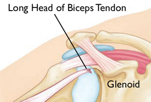
Biceps, as the name suggests, takes its origin from two heads – long head which arises from supraglenoid tubercle and passes over the humeral head within the capsule of the joint and the short head which arises from the coracoid process.

Near midshaft humerus, the two origins join to form a single muscle belly which then goes down and attaches to radial tuberosity below the elbow joint and blends with the bicipital aponeurosis, at cubital fossa.

Biceps muscle functions to flex elbow and supinate forearm. In addition, long head of biceps also acts as stabilizer of humeral head especially in external rotation and abduction.

biceps-tendon-rupture
Biceps Tendon Rupture, Image Credit: AAOS

Proximal biceps rupture results in a decrease of strength during shoulder flexion, elbow flexion, and forearm supination. Distal ruptures cause weakness of supination, elbow flexion, and grip strength.

The tears of biceps tendon could be partial or Complete tears.

Risks and Causes of Biceps Tendon Rupture

Older age, heavy overhead activities, shoulder overuse, smoking and corticosteroid medications and statins increase the risk of biceps tendon rupture.

Overuse can cause a range of shoulder problems, including tendonitis, shoulder impingement, and rotator cuff injuries. Having any of these conditions puts more stress on the biceps tendon, making it more likely to weaken or tear.

Most of the ruptures occur in individuals between the age of 40-60 years following chronic wear of the tendon. The causes of chronic wear are repetitive injury, chronic inflammation as in rheumatoid arthritis and rotator cuff injury.

Acute traumatic injury, heavy weightlifting, or injury during sporting activities may be the reason for rupture in younger patients.

Distal biceps tendon rupture is caused by chronic irritation on an irregular surface, such as in persistent cubital bursitis. Acute distal ruptures result from the forceful extension of the elbow from a flexed and supinated position.

Rapid flexion and adduction of the arm during elbow extension activities may cause short-head rupture though it is very rare.

Presentation of Biceps Tendon Rupture

Sudden, sharp pain in the upper arm, sometimes with an audible pop or snap is the general complaint in proximal biceps tendon rupture. There may be a history of pain in the upper arm prior to the incident especially on overhead activities or pain in the shoulder that may worsen at night.

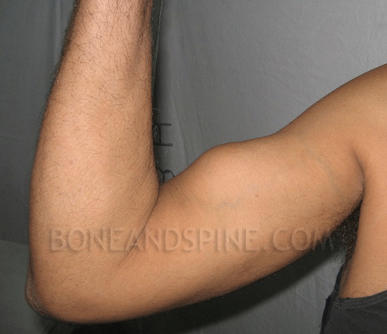
In contrast to this, there are patients who never experience anything and present with a palpable mass after rupture has occurred. A bulge in the upper arm above the elbow [Popeye Muscle see image below] may appear, with a dent closer to the shoulder.

Ruptured Distal Biceps Tendon

Tenderness may be present in course of the biceps muscle, especially in bicipital groove. Rotator cuff injuries and impingement may accompany a proximal biceps tendon rupture and shoulder joint should be examined for the range of motion and any weakness.

Speed test and Yergason sign, are used to rule out partial tears or biciptal tendonitis.

In distal biceps tendon ruptures, the symptoms are present near or at the elbow. A palpable gap is in the front of the elbow.

Imaging Studies

In most cases, proximal and distal ruptures can be detected on the basis of history and physical examination alone and imaging generally is not required.

However, AP and axillary views may reveal spurring or bony irregularities which increase the risk of biceps rupture.
Ultrasonography can reliably identify rupture of the proximal end of the long head of biceps but degenerative changes may not be identified easily.

Magnetic resonance imaging provides the best anatomic detail.

Treatment of Biceps Tendon Rupture

The pain of proximal biceps tendon rupture resolves with time. Resulting weakness is generally acceptable to patients with low demands. Because of this second attachment, many people can still use their biceps even after a complete tear of the long head.
Conservative management is considered appropriate for middle-aged or older patients and for those who do not require a high degree of supination strength in daily activities.

In the acute phase, ice for 20 minutes, several times a day should be applied. NSAIDs reduce pain and swelling. A sling is used to provide rest.

After the acute phase, the patient is put on flexibility and strengthening exercises.

Surgical treatment for a long head of the biceps tendon tear is rarely needed. Athletes or manual laborers who need strength may require surgery. Surgery can also be considered for partial tears where symptoms are not relieved with nonsurgical treatment.
Surgical repair consists of tenodesis and subacromial decompression. Successful surgery can correct muscle deformity and return strength and function.

Following surgery, the patient is gradually put on a range of motion exercises which are advanced to strengthening exercises.

Related

Spread the Knowledge
4
Shares
 
4
Shares
 4   

Filed Under: Hand and Upper Limb

About Dr Arun Pal Singh

Arun Pal Singh is an orthopedic and trauma surgeon, founder and chief editor of this website. He works in Kanwar Bone and Spine Clinic, Dasuya, Hoshiarpur, Punjab.

This website is an effort to educate and support people and medical personnel on orthopedic issues and musculoskeletal health.

You can follow him on Facebook, Linkedin and Twitter

Primary Sidebar

Browse Articles

Deformed pelvis oseomalcia

Osteomalacia Causes and Treatment

Osteomalacia is a  metabolic bone disease where defective mineralization results in bone soften due to a large amount of unmineralized osteoid. Osteomalacia (mollities ossium) is characterized by softening of the bones because of an accumulation of osteoid tissue. Osteoid is the bone matrix which is mineralized to form bone. Like rickets, osteomalacia also affects the […]

periosteal chondroma of femur

Juxtacortical Tumors or Surface Tumors of Bone

Juxtacortical tumors or surface tumors of bone are neoplasms [abnormal and excessive growth] arising from or just outside the cortex, and are composed of different histologic types. Some of the important juxtacortical tumors are Periosteal or juxtacortical chondroma Periosteal or juxtacortical chondrosarcomas Periosteal or juxtacortical  osteosarcomas Juxtacortical tumors are divided into 5 major categories Cartilaginous […]

a stretcher used for shifting spine patients

Spine Injuries – Presentation, Imaging and Treatment

Spine injuries are most commonly caused by motor vehicle injuries followed by fall and gunshot injury. Most of the spine injuries occur in cervical spine followed by thoracic and lumbar. Injuries of the cervical spine occur in more than half of all spine injuries in trauma patients. Most isolated thoracic and lumbar spine injuries are […]

Location of musculoskeletal tumors

Musculoskeletal Tumors – Classification, Diagnosis and Treatment

Musculoskeletal tumors are a rare and diverse group of tumors. Benign bone and soft tissue tumors are 100 times more common than malignant tumors. The musculoskeletal system is made of skin and subcutaneous tissue, lymphatic tissue, vascular structures, musculoaponeurotic tissue, neurological structures, bone, and joints. Musculoskeletal tumors of different histological types can present with similar […]

Parts of Shoe

Components of Orthopedic Shoes

Shoe serves a variety of functional and cosmetic purposes. For the individual with an orthopedic disorder, footwear can be selected to minimize pressure on sensitive, deformed structures. Shoes can be modified to redistribute weight toward pain-free areas to improve the comfort and function of the foot, limb, and trunk. The shoe is also is the […]

Fraser Classification of Floating Knee Injuries

Floating knee Injuries in Adults and Children

Floating knee refers to the fracture of ipsilateral [same side] fractures of tibia and femur. This makes knee unattached to tibia or femur, therefore name floating knee is given. The word floating knee was introduced for the first time by McBryde in 1965. Floating knee injuries may include a combination of diaphyseal, metaphyseal, and intra-articular […]

degenerative disc disease

Degenerative Disc Disease – Symptoms and Treatment

Degenerative disc disease is one of the most common causes of low back pain and neck pain. Degenerative disc disease describes the symptoms of pain and possibly radiating weakness or numbness stemming from a degenerated disc in the spine. The process is also termed as degenerative disc disorder. Degenerative disc disease can greatly affect the […]

© Copyright: BoneAndSpine.com
Manage Cookie Consent
The site uses cookies. Please accept cookies for a better visiting experience.
Functional Always active
The technical storage or access is strictly necessary for the legitimate purpose of enabling the use of a specific service explicitly requested by the subscriber or user, or for the sole purpose of carrying out the transmission of a communication over an electronic communications network.
Preferences
The technical storage or access is necessary for the legitimate purpose of storing preferences that are not requested by the subscriber or user.
Statistics
The technical storage or access that is used exclusively for statistical purposes. The technical storage or access that is used exclusively for anonymous statistical purposes. Without a subpoena, voluntary compliance on the part of your Internet Service Provider, or additional records from a third party, information stored or retrieved for this purpose alone cannot usually be used to identify you.
Marketing
The technical storage or access is required to create user profiles to send advertising, or to track the user on a website or across several websites for similar marketing purposes.
Manage options Manage services Manage vendors Read more about these purposes
View preferences
{title} {title} {title}
 

Loading Comments...