• Skip to primary navigation
  • Skip to main content
  • Skip to primary sidebar
  • Home
  • About
  • Newsletter/Updates
  • Contact Us
  • Policies

Bone and Spine

Orthopedic health, conditions and treatment

  • General Ortho
  • Procedures
  • Spine
  • Upper Limb
  • Lower Limb
  • Pain
  • Trauma
  • Tumors

Bipartite Patella

By Dr Arun Pal Singh

In this article
    • Relevant Anatomy
    • Classification of Bipartite Patella
    • How to Differentiate Fracture Patella and Bipartite Patella
    • Clinical presentation
  • Imaging
    • X-rays
    • MRI
    • Bone scan
  • Treatment of Bipartite Patella
    • Related

Bipartite patella is a congenital condition in which the patella is actually two separate bones instead of one and represents unfused or incompletely fused secondary ossification centers of the patella. Most of the people who have this condition are generally asymptomatic and most commonly. It may be confused with a fracture of the patella.

The incidence of occurrence of the bipartite patella is about 1% and is much more common in men than women [about 9 times]. It occurs bilaterally in about 50% of cases. Association with nail-patella syndrome has been noted.

Xray of Bipartite Patella
Xray of Bipartite Patella, fragment marked with the arrow, Image modified from Radiopaedia

Relevant Anatomy

The patella is the largest sesamoid bone which starts ossifying at an age of 3-4.5 years. Accessory ossification center appears between 8-12 years and fuses by puberty. Failure of fusion leads to the presence of a separate fragment. This is termed as bipartite patella.

Classification of Bipartite Patella

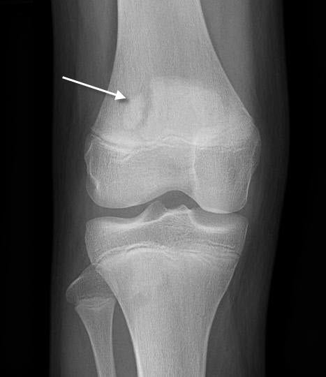
Saupe Classification describes 3 types of bipartite patella depending on location of separate fragment.

Classification of Bipartite Patella
Type I

Inferior pole

Type II

Lateral margin

Type III

Superolateral pole

How to Differentiate Fracture Patella and Bipartite Patella

When compared with patellar fracture, the bipartite patella is

  • Located superolaterally
  • Have rounded borders
  • Contralateral knee x-ray may have similar findings because bipartite patella is bilateral in 50% cases

Clinical presentation

A bipartite patella is usually an incidental finding. Only 2% of patients with the condition experience symptoms. It may cause anterior knee pain, especially after trauma, sports injury, or overuse. Direct trauma can occur due to fall or kick to the knee. Indirect trauma can occur due to repetitive injury as in cycling, hill climbing.

The pain gets aggravated by squatting, jumping or climbing stairs. A sensation of giving away may also be felt.

Causes of knee pain in bipartite patella are

  • Injury to the fibrocartilaginous zone between the main patella and accessory fragment
  • Fibrocartilaginous zone does not heal leading to persistent pain
  • Repeated vastus lateralis force applies to fragment resulting in separation and nonunion

Physical examination may reveal a larger than normal patella with localized tenderness over accessory fragment. A hematoma or patellar prominence may be felt.

Imaging

X-rays

AP knee radiograph is the best view to visualize. Another view that would reveal bipartite patella is skyline view. Xray of the opposite knee is done to check the presence of the condition in the other knee.

MRI

MRI can reveal if the pain is attributable to the bipartite patella. It may show edema around the fragment in symptomatic patients.

Bone scan

It is rarely needed in patients with suspicion for bipartite patella and inconclusive radiographs. Bone scan shows increased uptake along the fragment.

Treatment of Bipartite Patella

Symptomatic bipartite patella usually improves with nonoperative treatment. Nonoperative treatment includes rest, immobilization with the knee braced in 30° of flexion, NSAIDS, and physical therapy [isometric stretching exercises of the quadriceps muscle in extension].

Local corticosteroid injection also eases out the symptoms.

Operative treatment consists of excision of the accessory fragment and is indicated in

  • No improvement on nonoperative treatment even after six months.
  • The articular surface of the fragment is irregular.

Fixation may be required in large fragments.

Related

Spread the Knowledge
10
Shares
 
10
Shares
19   

Filed Under: Knee

About Dr Arun Pal Singh

Arun Pal Singh is an orthopedic and trauma surgeon, founder and chief editor of this website. He works in Kanwar Bone and Spine Clinic, Dasuya, Hoshiarpur, Punjab.

This website is an effort to educate and support people and medical personnel on orthopedic issues and musculoskeletal health.

You can follow him on Facebook, Linkedin and Twitter

Primary Sidebar

Browse Articles

Fibula bone different views

Fibula Bone Anatomy

The leg has two bones – tibia and fibula. The fibula is very thin bone compared to the tibia and can be expended except for small length distally. This makes it usable for various orthopedic procedures. The fibula is homologous with the ulna. The fibula is a located on the lateral side of the tibia, […]

Calcium Homeostasis is complexely egulated

Calcium Homeostasis – Physiology and Disorders

Calcium homeostasis refers to the regulation of the concentration of calcium ions in the extracellular fluid. An adult body contains about one kilogram of calcium. It is the fifth most abundant element in the human body. Calcium takes part in a wide range of biologic functions including bone mineralization and. Calcium is mainly provided by […]

antitubercular chemotherapy doses

Antitubercular Chemotherapy in Musculoskeletal Tuberculosis

Antitubercular chemotherapy is the mainstay of the treatment of osteoarticular tuberculosis. It is complemented by rest, nutritional support and splinting, as necessary. The drugs and regimens are fundamentally similar to those for pulmonary TB. But there is a lack of consensus on the appropriate duration of treatment. Overview of Osteoarticular Tuberculosis The spine is probably […]

salter pelvic osteotomy

Pelvic osteotomies – Indications and Types

Pelvic osteotomies are done to stabilize the hip and prevent early osteoarthritis in cases of acetabular dysplasia [Altered congruency of the acetabulum resulting from its maldevelopment]. The acetabulum is the large cup-shaped cavity on the anterolateral aspect of the pelvis that articulates with the femoral head to form the hip joint. Persistent acetabular dysplasia can lead […]

Lacerated Wound On Ankle

Classification of Wounds

Classification of wounds can be done in many ways. Classification of Wounds Based on the Thickness of the wound Superficial Wounds These involve only the epidermis and the dermis up to the dermal papillae. Partial-thickness Wounds Partial thickness wounds involve skin loss up to the lower dermis. Full-thickness Wounds Full thickness wounds involve the skin […]

Treatment algorithm of Pott's paraplegia

Pott’s Paraplegia or Spinal Tuberculosis with Neural Involvement

Spinal tuberculosis  with neural deficit can occur in the lesions when disease comrpresses the cord. Potts paraplegia is term for this deficit in TB spine Spinal tuberculosis  is the most common form of skeletal tu­berculosis accounting for 50 percent of osteoarticular tuberculosis. Neural compression in spinal tuberculosis may lead to tetraparesis-tetraplegia or paraparesis-paraplegia. Pathology of […]

tibia fibula interosseous membrane

Tibiofibular Joint Anatomy

Tibiofibular joints are articulations between tibia and fibula. Superior tibiofibular joint is articulation between head of fibula and upper tibia. At ankle, the articulation between tibia and fibula is called inferior tibiofibular joint. Two bones are connected by interosseous membrane which is also sometimes called middle tibiofibular joint. Superior Tibiofibular Joint The superior tibiofibular joint […]

© Copyright: BoneAndSpine.com
Manage Cookie Consent
The site uses cookies. Please accept cookies for a better visiting experience.
Functional Always active
The technical storage or access is strictly necessary for the legitimate purpose of enabling the use of a specific service explicitly requested by the subscriber or user, or for the sole purpose of carrying out the transmission of a communication over an electronic communications network.
Preferences
The technical storage or access is necessary for the legitimate purpose of storing preferences that are not requested by the subscriber or user.
Statistics
The technical storage or access that is used exclusively for statistical purposes. The technical storage or access that is used exclusively for anonymous statistical purposes. Without a subpoena, voluntary compliance on the part of your Internet Service Provider, or additional records from a third party, information stored or retrieved for this purpose alone cannot usually be used to identify you.
Marketing
The technical storage or access is required to create user profiles to send advertising, or to track the user on a website or across several websites for similar marketing purposes.
Manage options Manage services Manage vendors Read more about these purposes
View preferences
{title} {title} {title}
 

Loading Comments...