Last Updated on January 2, 2024
Blood supply of neck of femur is derived from vessels supplying part of the femur. The blood supply of head of the femur is also contributed by these vessels. Blood supply of neck of femur is important to understand because disruption of the blood supply during trauma or surgery can lead to avascular necrosis of the femoral head. It helps to gauge the risk of avascular necrosis and plan the surgical dissection to preserve the blood supply.
Relevant Anatomy
The femoral head is the most proximal part of the femur and is connected by the femoral neck to the rest of the femur. The femoal head forms the ball of the hip joint that articulates with the socket formed by the acetabulum.
The head is almost spherical and has a medial depression known as the fovea capitis femoris which serves as an attachment point for the ligamentum teres.
Many hip muscles stabilize the hip and allow various movements like flexion, extension, abduction, adduction, internal rotation, and external rotation.
Blood Supply of Neck of Femur and Head of Femur
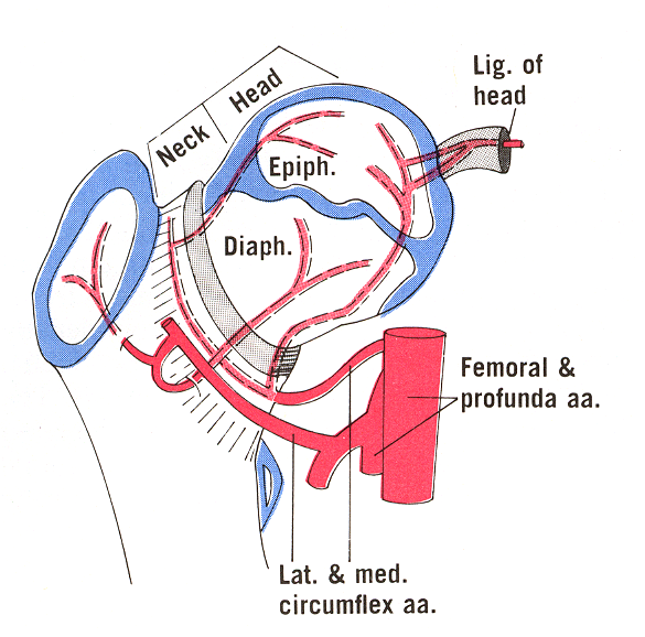
The majority of the blood supply to the neck of femur and head of femur comes from the medial and lateral circumflex arteries. These circumflex arteries are branches of the profunda femoris, which in turn is a branch of the femoral artery.
The medial and lateral circumflex form a ring around the neck of the femur by anastomosis and from that small arteries travel to supply the head.
The foveal artery or the artery of the ligamentum teres runs within the ligament and contributes to the head of femur supply in children.
Details of Vascular Arrangement
Extracapsular ring (At the base of femoral neck)
It is formed by following vessels
- Medial circumflex femoral artery (MCFA) – posteriorly
- Lateral circumflex femoral artery (LCFA) – anteriorly
- Minor contribution from superior and inferior gluteal arteries
- Anastomoses
- Trochanteric anastomosis- centered on trochanteric fossa
- Between the superior gluteal artery and medial/lateral circumflex femoral arteries.
- Trochanteric anastomosis- centered on trochanteric fossa
-
- Cruciate anastomosis
- Between the inferior gluteal artery and the medial circumflex femoral artery
- Cruciate anastomosis
- Weathersby anastomosis
- Infra-acetabular region
- Superior gluteal artery
- Inferior gluteal artery
- Lateral femoral circumflex artery
- Oburator arteries
- Infra-acetabular region
Retinacular Vessels
Ascending cervical vessels arise from the extracapsular ring and travel proximally under the hip capsule and continue proximally along the neck deep to the synovial membrane toward the femoral head.
These arteries are known as retinacular arteries and are divided into three groups
- Posterior inferior & posterior superior (from the medial femoral circumflex artery)
- Anterior (from the lateral femoral circumflex artery)
At the margin of the articular cartilage on the surface of the neck of the femur, these vessels form the second ring called subsynovial intra-articular ring from which arise epiphyseal arteries.

Retinacula of Weitbrecht are folds formed by reflections or continuations of the synovial membrane combined with fibrous sheath from the capsular wall. These are three in number and carry blood vessels within. The three retinacula are
- Medial or inferior
- Lateral or superior
- Anterior
Medial is the most constant and anterior is seen in only 40 percent.
Subsynovial intracapsular ring
Also called circulus articuli vasculosus of Hunter, these are found at the junction of articular surface of the femoral head with neck)
The ring is stronger on medial and lateral aspects than anterior and posterior.
It can be incomplete increasing the predilection of Legg-Calve-Perthes disease in male gender.
The deep branch of MFCA gives rise to the lateral epiphyseal artery which supplies the majority of the femoral head including the weight-bearing surface is the most important and is particularly prone to injury.
Artery of ligamentum teres
It is a branch of obturator artery and enters through fovea capitis). Occasionally it may arise from medial circumflex femoral artery.
It continues as medial epiphyseal artery.
Epihyseal and Metaphyseal Arteries
These arteries supply epiphyses and metaphyses during the growth phase. These anastomose after the closure of physis and participate in intraosseous blood supply of neck of femur and head.
Intraosseous Blood Supply of Neck of Femur
The intramedullary branches of nutrient [arise from upper perforating arteries of the profunda femoris], metaphyseal [Arise from medial circumflex and extracapsular arterial ring and subsynovial ring] and epiphyseal vessels [Arise from subsynovial ring] feed both marrow and cortical bone. If the fracture of neck of femur is complete, this blood supply of neck of femur and head gets disrupted.
Artery of Ligamentum Teres
It is a branch of the medial circumflex femoral artery and supplies the head of the femur through ligamentum teres. It also forms the medial epiphyseal vessels. Only a small & variable amount of the femoral head is supplied by the artery of ligamentum teres.
Note:
- Medial femoral circumflex artery arises from the posteromedial aspect of the deep femoral artery
- Lateral femoral circumflex usually arises from the lateral side of the deep femoral artery
Clinical Significance of Blood Supply of Neck of Femur
Legg-Calve-Perthes Disease
This refers to the idiopathic necrosis of proximal femoral epiphysis in children. There is a disruption of blood supply to the head of the femur followed by revascularization over 2-5 years.
The prognosis is good though some children have persistent deformed head which increases the risk of early osteoarthritis.
Femoral Head Fractures
These are rare fractures and are almost always associated with hip dislocation. They require anatomical reduction and fixation as the injury disrupts the blood supply of head of femur.
Avascular Necrosis of Head of the Femur
This is often bilateral and has systemic causes. There is a disruption of blood supply and ischemic death of the bone cells follows.
Progression of the condition may lead to head deformity and degenerative changes.
References
- Zlotorowicz M, Czubak-Wrzosek M, Wrzosek P, Czubak J. The origin of the medial femoral circumflex artery, lateral femoral circumflex artery and obturator artery. Surg Radiol Anat. 2018 May;40(5):515-520. [Link]
- Gautier E, Ganz K, Krügel N, Gill T, Ganz R. Anatomy of the medial femoral circumflex artery and its surgical implications. J Bone Joint Surg Br. 2000 Jul;82(5):679-83. [Link]
Do leave your feedback on this article about blood supply of neck of femur.