Last Updated on December 3, 2023
The blood supply of spinal cord and the vertebral canal is very important from surgical and clinical point of view in addition to its vital importance.
The spinal cord is located within the vertebral canal. The blood supply of spinal cord is a complex arrangement of arteries and veins and is responsible for adequate circulation of the cord.
In this article, the blood supply of spinal cord is discussed along with its variations and important implications.
Relevant Anatomy of Vertebral Canal and Spinal Cord
The spinal cord begins at the base of the foramen magnum, a large hole in the base of the skull and extends to conus medullaris [terminal end of the spinal cord]. The conus medullaris is generally between spinal vertebrae L1-L2 but can range from T12 to between L2-L3 in different individuals.
Three membranes, called meninges surround the spinal cord
- Dura mater is the outermost
- Arachnoid mater is the middle
- Pia mater is the innermost
The meninges extend down further than the spinal cord and house the exiting lower nerve roots [filum terminale] for some distance. The subarachnoid space terminates around the S2 vertebrae.
The spinal cord is contained within the vertebral canal. The vertebral canal is formed by the stacking of vertebrae from cervical to the sacral region by
- 7 cervical vertebrae
- 12 thoracic
- 5 lumbar
- 5 sacral
The vertebrae are load-bearing structures. the main load is borne by the body of the vertebra. Between two vertebrae lies the cushioning structure called the intervertebral disc.
The body of the vertebra is the main weight-bearing structure and is separated by the intervertebral discs (which allow cushioning). The vertebral size increases as we move downward till L5.
Also read: Anatomy of vertebral canal
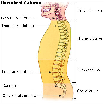
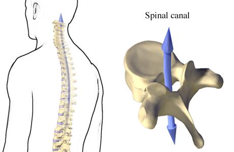
Image Credit: Alberta
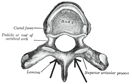
The vertebral arch forms the posterior and lateral aspects of the vertebrae and has a central lumen that houses the spinal cord.
The arch is made of pedicles and laminae. The pedicles attach the arch to the body. The laminae are extensions from the pedicle that extend and converge to meet at the midline. Spinous processes arise from that junction.
At each side of the pedicle, superior articular processes and inferior articular processes are located. Superior articular processes of each vertebra articulate with the inferior articular process of the vertebra on top and inferior ones articulates with superior articular processes of the vertebra below.
The inferior and superior articular processes via which the vessels and nerves traverse.
Transverse processes are formed at the junction between the pedicle and laminae and face posterolaterally.
Blood vessels form from the mesoderm. During development as the nerve roots that will connect to the spinal cord
Detailed Blood Supply of Spinal Cord
The blood supply of spinal cord is provided by different sources which vary with different regions of the spine.
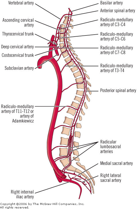
The blood supply of spinal cord is mainly dependent on three vessels. These are namely
- Anterior median longitudinal arterial trunk or anterior spinal artery
- Supplies anterior two-thirds of the cord
- Formed by branches from vertebral arteries
- Posterolateral trunks or posterior spinal arteries
- Paired arteries
- Supply the posterior one-third of the spinal cord
There are various interconnections between these supplies
- Inferiorly, the cruciate anastomosis of the conus medullaris
- Pial anastomoses called the arterial vasocorona
- Encircles the cord and supplies the peripheral lateral aspect of the spinal cord.
These longitudinal arterial trunks are most considerable in size in the cervical and lumbar regions and the size is much smaller in the thoracic region.
Generally speaking, the blood supply of spinal cord in the thoracic area is poor as compared to the cervical and lumbar region. The blood supply of the spinal cord is poorest between levels T4 and T9.
However, these are not the sole vessels on which the blood supply of the spinal cord is dependent. There are many other reinforcing vessels. A list of other vessels supplying the spinal cord is as follows-
- Radicular arteries of the cord
- Vertebral Arteries
- Posteroinferior cerebellar arteries
- Segmental Arteries
- Ascending cervical arteries
- Deep cervical arteries
- Posterior intercostal arteries
- Subcostal arteries
- Lumbar arteries
- Lateral sacral arteries
Major arterial supply of the cord is discussed below
Anterior Spinal Artery and Posterior Spinal Arteries
The anterior spinal artery and two posterior spinal arteries are the main suppliers of blood to the cord.
The anterior spinal artery is formed by the vertebral arteries. Vertebral arteries originate from the first part of the subclavian artery.
As they ascend, they pass through the transverse foramen of C1 through C6 and then through the foramen magnum to become the basilar artery.
The vertebral arteries are given off as branches before basilar arteries. The anterior spinal artery is a branch of the vertebral artery and travels causally down the spinal cord through the anterior sulcus.
The posterior spinal arteries can be branches of the posterior inferior cerebellar artery or the pre-atlantal vertebral arteries. These also travel which also travel caudally down the spinal cord through the two posterior sulci.
Thus anterior spinal artery is anterior to the cord and posterior spinal arteries lie posterior to the cord.
The anterior spinal arteries provide the blood to the anterior two-thirds of the spinal cord and the posterior spinal arteries to the posterior one-third.
Other Important Vessels Supplying the Spinal Cord
Radicular Arteries of The Cord
Also called medullary feeder arteries, these reinforce the longitudinal arterial channels.
These vary from 2-17 on the anterior side and 6-25 on the posterior side.
The vertebral arteries supply 80% of feeder arteries in the neck. In thoracic and lumbar areas the aorta gives rise to these vessels. In the sacral area, iliolumbar arteries, fifth lumbar arteries, middle sacral arteries, and lateral sacral arteries are important sources.
Vertebral and Posteroinferior Cerebellar Arteries
These form an additional important source of blood supply in the cervical region.
Segmental Arteries
Segmental arteries are at every level of the spine and occur in pairs. These supply both spinal and extraspinal tissues.
They originate from vertebral arteries in the neck, aorta in the thoracolumbar area, and lateral sacral artery in the sacral region.
In a few people ascending pharyngeal artery is an important source of segmental arteries.
The segmental arteries branch profusely at the intervertebral foramen and a second anastomosis occurs within the spinal canal in the extradural region.
This anastomosis forms a rich network of vessels and allows to ligation of segmental arteries without affecting the circulation of the cord.
Artery of AdamKiewz
It is the largest feeder of the lumbar part of the spinal cord and forms an important component of blood supply of spinal cord. It is on the left side between the T9-T11 vertebrae in almost 80% of people.
The artery of Adamkiewicz takes its origin mostly from the left posterior intercostal artery between vertebra T9 and T12.
Immediately, it extends through the intervertebral foramen of the corresponding spinal level and ascends the anterior surface of the spinal cord for about two vertebrae or slightly more and then undergoes a hairpin curve and anastomoses with the anterior spinal artery.

Venous Drainage of Spinal Cord
Venous drainage of the spinal cord is not as clearly defined as arterial circulation and can be highly variable.
Generally speaking, there are two sets of vessels
- Veins of spinal cord
- Venous plexus of Batson
The veins of the spinal cord are small veins and form a small component of the venous system. they drain into Batson plexus.

Batson plexus is a large and complex venous channel communicating directly with inferior and superior venae cavae. It extends from the base of the skull to the coccyx. The Batson plexus has three components
- Extradural venous plexus
- Extravertebral venous plexus
- Veins to osseous structures of the spine
The longitudinal venous trunks are called anterior and posterior venous channels.
Clinical Significance of Blood Supply of Spinal Cord
The knowledge of arterial circulation is important during surgical procedures. As for as possible the blood supply of spinal cord should be preserved.
As the venous system communicates directly with large vessels, this interconnection allows metastatic spread from the pelvis to the vertebral column.
Video on Blood Supply of Spinal Cord
The following video by Learning Anatomy would help you to revise the concepts of spinal cord blood supply
References
- Boll DT, Bulow H, Blackham KA, Aschoff AJ, Schmitz BL. MDCT angiography of the spinal vasculature and the artery of Adamkiewicz. AJR Am J Roentgenol. 2006 Oct;187(4):1054-60.
- Martirosyan NL, Feuerstein JS, Theodore N, Cavalcanti DD, Spetzler RF, Preul MC. Blood supply and vascular reactivity of the spinal cord under normal and pathological conditions. J Neurosurg Spine. 2011 Sep;15(3):238-51.