Last Updated on February 19, 2025
Bone mineralization is the process of laying down minerals on a matrix of the bone. Normal bone is composed of 50 to 70% mineral, 20 to 40% organic matrix, 5 to 10% water, and <3% lipids. Calcium and phosphorus are chief minerals found in the bone along with a small amount of carbonate, and magnesium.
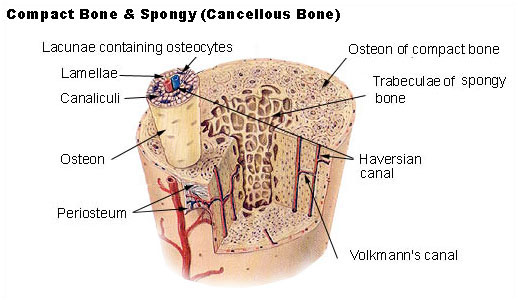
The mineral content of bone is mostly hydroxyapatite [Ca10(PO4)6(OH)2]. Bone mineralization is a well-regulated process in which crystals of calcium phosphate are produced by bone-forming cells [osteoblasts] and are laid down in precise amounts within the fibrous matrix.
Any defect in the process at any level can lead to too little or too much mineral content.
The process of mineralization [also called calcification] takes place throughout the entire life.
The terms ‘calcification’ and ‘mineralization’ are used interchangeably. However, mineralization is most often used in the context of bone whereas calcification refers to the accumulation of calcium salts in a tissue and is often pathological.
Bone Formation
Bone is a composite tissue comprised of both organic and mineral components, specialized cells, and water. The bone formation begins with the synthesis and deposition of the extra-cellular matrix or osteoid followed by mineralization. The osteoid is formed primarily of type I collagen. It also contains non-collagenous proteins, growth factors, and glycoproteins.
The mineral component of bone is hydroxyapatite (Ca10(PO4)6(OH2)). The mineral component is formed by inorganic phosphate (PO4)3- or also written as Pi) and calcium (Ca2+) ions.
The cells of bone responsible for formation are osteoblasts. These are derived from mesenchymal stromal cells.
The Process of Bone Mineralization
The exact mechanisms by which mineralization occurs remain unclear. There are two main theories
- Matrix vesicle-mediated: Matrix vesicles are extracellular and are secreted into the osteoid by osteoblasts and are thought to facilitate mineralization by accumulation of Ca2+ and P ions.
These ions combine to produce a calcium-phosphate intermediate that leads to the formation of hydroxyapatite. As the size of the crystal enlarges, the membrane of the vesicle ruptures and the crystal is released into the extracellular environment.
Subsequent growth of the hydroxyapatite crystals is then dependent on the extracellular concentrations of Ca2+ and Pi and on the levels of local mineralization inhibitors - Collagen template: Bone mineral is initially deposited in “hole” zones between the ends of collagen fibrils of the matrixTemplate-mediated mineralization proposes that the 40 nm hole regions within the collagen fibrils allow Ca2+ and Pi to accumulate, precipitate, and form hydroxyapatite crystals.
It is also proposed that both mechanisms may occur.
Regulators of Bone Mineralization
Osteoblasts form and secrete inorganic and organic constituents of the extracellular matrix. When matrix maturation occurs, there is an expression of alkaline phosphatase and several proteins, including osteocalcin, osteopontin, and bone sialoproteins.
The process of bone mineralization is tightly regulated by a variety of local and systemic factors so that both under and over-mineralization does not occur.
These are
- Pyrophosphate (PPi): Two molecules of inorganic phosphate join together to form pyrophosphate (PPi). Extracellular pyrophosphate potently inhibits the ability of Ca2+ to crystallize with phosphate ion or Pi to form hydroxyapatite and also prevents crystal growth.
- Pyrophosphatases/phosphodiesterases: These hydrolyze the phosphodiester bond in nucleotide triphosphates like ATP to generate PPi
- Tissue non-specific alkaline phosphatase (TNSALP): It can hydrolyze PPi to generate Pi and thus favors mineralization. It is antagonistic to the action of phosphodiesterases like NPP1.
- PHOSPHO1: This enzyme hydrolyses phosphoethanolamine and phosphocholine to release Pi. It contributes to the initiation of mineralization.
- Non-collagenous proteins (NCPs): These are synthesized by osteoblasts and account for 10 percent of the extracellular matrix. Important ones are matrix gla protein (MGP), osteocalcin (OCN) and osteopontin (OPN). which can regulate the process. MGP and OPN bind hydroxyapatite and prevent crystal growth while OPN’s role is not well elucidated.
- Extracellular nucleotides: ATP, ADP, and UTP can modulate osteoblast differentiation, proliferation, and function
- Vitamin D: This along with parathyroid hormone plays a key role in the regulation of Ca2+ and Pi homeostasis.
- Ion transporters: Chloride-proton antiporters such as CIC3 and CIC5 work to remove H+ ions formed during crystallization so that pH is maintained.
Feedback Loops of Bone Mineralization
Bone mineralization is controlled at three levels by delicate feedback loops
- Systemic hormones – Vitamin D, Parathyroid etc
- Bone cells (osteoblasts, osteoclasts, and osteocytes)
- Cell products
The mineral ions Ca2+ and inorganic phosphate [Pi], and PPi play a central role in controlling the bone mineralization process.
An increase in extracellular PPi leads to a decrease in hydroxyapatite whereas a decrease in PPi results in increased skeletal mineralization.
Bone mineralization is dependent on a tight local balance between extracellular levels of Pi and PPi.
The skeleton acts as a reservoir for the storage of both Ca2+ and Pi.
Regulation of blood Ca2+ and Pi levels involves bone, kidney, and intestine and hormones like parathyroid, vitamin D3, FGF23
Parathyroid hormone is secreted in response to low serum Ca2+ levels. It regulates bone remodeling directly via actions on osteoblasts and indirectly by actions in other tissues (such as increasing the formation of active vitamin D3 in the kidney).
Vitamin D plays an indirect role in stimulating the mineralization of unmineralized bone matrix. It stimulates the intestinal absorption of calcium and phosphorus to achieve enough calcium concentration. It also promotes the differentiation of osteoblasts and stimulates osteoblast expression of bone-specific alkaline phosphatase, osteocalcin, osteonectin, etc.
Parathyroid hormone can act on osteoblasts to directly regulate the expression of genes important in bone remodeling (e.g. RANKL)
FGF23 is released by osteocytes and osteoblasts in response to high Pi levels. It acts to inhibit Pi reabsorption in the kidneys.
When the bone mineralization is about to end approximately 60–90% of osteoblasts are estimated to die and many of the remaining cells undergo a terminal differentiation into an osteocyte.
References
-
Murshed M. Mechanism of Bone Mineralization. Cold Spring Harb Perspect Med. 2018 Dec 3;8(12):a031229. doi: 10.1101/cshperspect.a031229. Erratum in: Cold Spring Harb Perspect Med. 2020 Aug 3;10(8):a040667. [doi]
-
Bonucci E. Bone mineralization. Front Biosci (Landmark Ed). 2012 Jan 1;17(1):100-28. doi: 10.2741/3918.