• Skip to main content
  • Skip to primary sidebar
  • Skip to footer
  • General Ortho
  • Procedures
  • Spine
  • Upper Limb
  • Lower Limb
  • Pain
  • Trauma
  • Tumors
  • Newsletter/Updates
  • About Us
  • Contact Us

Bone and Spine

Orthopedic health, conditions and treatment

Bones in Human Body and Their Function

By Dr Arun Pal Singh

In this article
    • The Human Skeleton
    • Region-wise Bones in Human Body
      • Skull [28 bones]
      • Spine
      • Throat
      • Thorax
      • Upper limbs
      • Hand & Wrist
      • Pelvis (2 bones)
      • Lower Limbs (60 bones, 30 each side)
      • Feet
    • Functions of Bones or Human Skeleton
      • Support and Locomotion
      • Protection of Underlying Structures
      • Making Blood Cells
      • Storage
      • Immunity

The human skeleton forms the main supporting framework of the body. The skeleton is comprised of an arrangement of different bones in human body for the more efficient production of movements by the attached muscles.

There are 206 bones in human body (The count may vary to 208 depending on whether sternum is counted as three bones instead of one bone with three parts.

At birth, there are 270 bones in the human body. The count decreases to 206 as many of the bones in the childhood fuse with growth.

Another important point to make is that the coccyx and sacrum are fused in adults but these are not counted single bone but as the number of individual vertebrae they have. Thus, the count will decrease further if sacrum and coccyx are considered as single bone each.

Many small accessory bones, such as some sesamoid bones, are not included in this count.

Let us familiarize ourselves with bones in our body. We start from the top and move down.

It is important to understand normal structures and functions to locate abnormality. Only then we can recognize abnormal functions. Our skeleton is our basic structure on which everything else is laid.

The Human Skeleton

skeleton showing joints and bones in human body

The human skeleton is divided into

  • Axial skeleton
  • Appendicular skeleton

The axial skeleton is the central part of the skeleton and comprises of 80 bones that form

  • Head Spine
  • Chest
  • head

The appendicular skeleton refers to the limbs and comprises of 126 bones in the form of

  • Shoulder girdle
  • Arms
  • Pelvic girdle
  • Legs

Thus, there are a total of 206 bones in human body across the entire skeleton.

Region-wise Bones in Human Body

Skull [28 bones]

Skull Bones

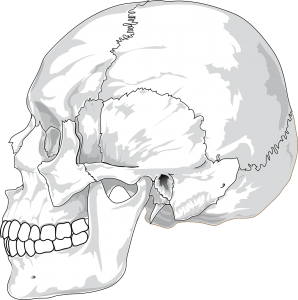
Skull is formed by cranium and mandible. Cranium is further composed of multiple bones that are united to each other. The lines of the union are called sutures. these sutures are lax in children to allow brain growth. Mandible constitutes our lower jaw that we used the whole day for speaking, eating or gesturing.

There are 22 bones in the skull and 6 in the middle ear, making a total of 28 bones. Skull has 8 bones in cranium and 14 in the face

Cranial bones (8)

  • Occipital bone (1)
  • Parietal bones (2)
  • Frontal bone (1)
  • Temporal bones (2)
  • Sphenoid bone (1)
  • Ethmoid bone (1)

Sphenoid bone and ethmoid bone are considered as facial bones by some authors.

Facial bones (14)

  • Nasal bones (2)
  • Maxillae or upper jaw (2)
  • Lacrimal bone (2)
  • Zygomatic bone or cheekbones (2)
  • Palatine bone (2)
  • Inferior nasal concha (2)
  • Vomer (1)
  • Mandible (1)

Middle ears (6 bones)

Each ear has 3 bones making a total of 6 bones in both ears.

  • Malleus (2)
  • Incus (2)
  • Stapes (2)

Spine

This is formed by multiple vertebrae which are named according to area

vertebral column
Vertebral Column

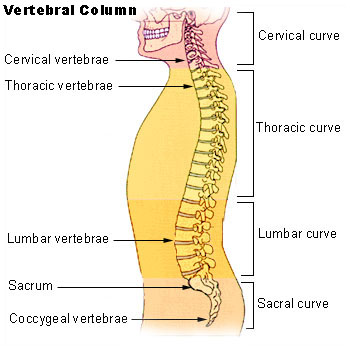
A child has 33 vertebrae. In adults, sacral and coccygeal vertebrae fuse. The number of bones each region has are

  • Cervical vertebrae (7 bones)
  • Thoracic vertebrae (12 bones)
  • Lumbar vertebrae (5 bones)
  • Sacral vertebrae (5 bones at birth but fuse later)
  • Coccygeal vertebrae (4 bones)

If sacrum and coccyx is considered as a single bone, the count of bones in the vertebral column would be 26
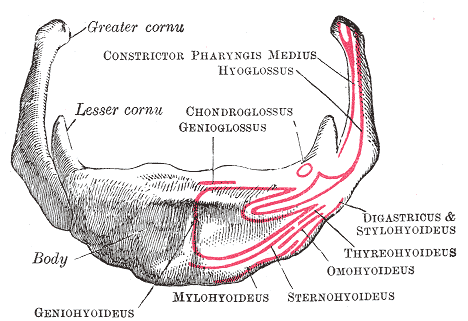
Throat

The hyoid bone is present in the throat and serves as support to cartilages of trachea or windpipe. It also serves as attachments to various muscles that help in movement of the trachea.

hyoid bone

Thorax

Thorax Bones

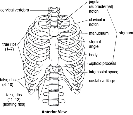
Thorax is a cage-like structure that functions to protect vital structures that it contains like lungs, heart etc. it is formed by ribs on either side which are connected to the sternum in front and to vertebrae on back.

  • Sternum (1)
  • Ribs (24, 12 on each side)

Upper limbs

Upper limbs include shoulder girdle, arm, forearm, wrist and hand. Upper limbs contain 64 bones, 32 on each side. Below the numbers are given for single extremity.

Shoulder Girdle

Bones of Shoulder Girdle

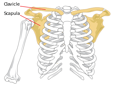
Shoulder Girdle is an area of the shoulder that is formed calvicle (1) and scapula (1) articulating with humerus (1).

  • Clavicle : It is a bone that connects the thoracic cage to upper limbs and also functions for the transmittal of forces.
  • Scapula: It is a complexly structured bone that articulates with the clavicle, thorax and takes part in the formation of the shoulder joint.

Arm

The humerus is the only bone that is in the arm.

Humerus bone

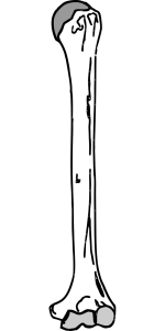
The bone that connects the shoulder to the elbow. This bone is normally cylindrical but flares when it comes near the elbow and becomes flat. It flares on either side to form ridges called condyles.

Ulna and Radius (1,1)

Ulna is the bone that forms elbow joint by articulating with the humerus. This is the bone that protrudes on back of elbow on flexion. It can be felt on the aspect of the forearm on side of the little finger.

Radius and Ulna

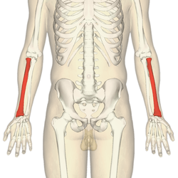
Radius is [depicted red in image] the bone on other side of forearm i.e. on side of the thumb. It forms a joint with ulna on the upper side. The part that takes part in this joint formation is called the head of the radius. This joint allows twisting movements of the elbow called pronation and supination.

Hand & Wrist

The hand is quite a complex structure and requires a separate discussion. Here are the names of bones. We would discuss them in detail in carpal anatomy and hand anatomy.

Hand wrist bones

hand x-ray

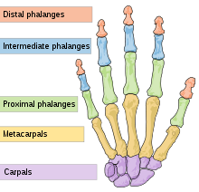
  • Carpals or Wrist Bones (8)
    • Scaphoid [A]
    • Lunate[B]
    • Triquetrum [C]
    • Pisiform [D]
    • Trapezium [E]
    • Trapezoid [F]
    • Capitate [G]
    • Hamate [H]
  • Metacarpals (4)
  • Phalanges (14)
    • Proximal phalanges (5)
    • Intermediate phalanges (4)
    • Distal phalanges (5)

*Thumb has only two phalanges – proximal and distal
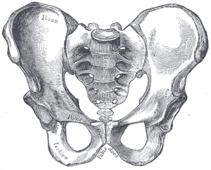
Pelvis (2 bones)

Pelvis or pelvic girdle is formed by many bones on either side which on one hand are connected to the spine and on the other side to lower limbs. Pelvis functions to transmit the weight to lower limbs and also protect the vital structures like rectum and bladder.

Ilium, pubis, and ischium are three bones that unite to form hip bone on each side. These articulate with sacroum and coccyx column to complete pelvis. Thus there is a total of 2 hip bones.

More on pelvic anatomy

Pelvic Bones

Lower Limbs (60 bones, 30 each side)

The structure of the lower limb is quite similar to upper limbs with modifications done for weight-bearing purposes. The lower limb contains 30 bones each side.

There are a total of 60 bones in the lower limbs.

  • Femur (2 bones, 1 each side)
  • Patella or kneecap (2 bones, 1 each side)
  • Tibia (2 bones, 1 each side)
  • Fibula (2 bones, 1 each side)
  • Foot (52 bones in total, 26 per foot)


Bones of Lower Limb

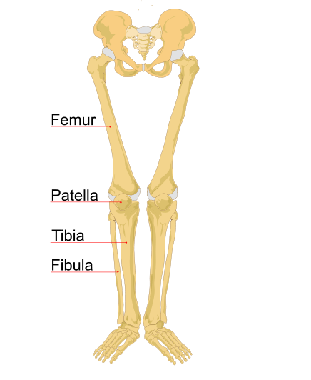
Femur, tibia, and fibula are long bones that form the lower limb. The femur is bone of thigh whereas the other two bones are present in the leg. Another important bone called patella is present on the anterior side of the knee joint and is commonly known as the kneecap.

Feet

Foot bones Tarsal bones

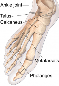
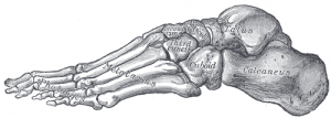
The foot contains 52 bones, 26 per foot.

  • Tarsals (14 bones, 7 each foot)
    • Calcaneus (2 bones, 1 each side)
    • Talus (2 bones, 1 each side)
    • Navicular bone (2 bones, 1 each side)
    • Medial cuneiform bone (2 bones, 1 each side)
    • Intermediate cuneiform bone (2 bones, 1 each side)
    • Lateral cuneiform bone (2 bones, 1 each side)
    • Cuboidal bone (2 bones, 1 each side)
  • Metatarsals (10 bones, 5 each side)
  • Phalanges ( 28 bones, 14 each side)
    • Proximal phalanges (10 bones, 5 each side)
    • Intermediate phalanges (8 bones, 4 each side)
    • Distal phalanges (10 bones, 5 each side)

Bones can be classified by many methods. Here we discuss different types of bones one by one. Bone classification can be done by various parameters.


Functions of Bones or Human Skeleton

Support and Locomotion

Bones in  human body provide basic structural shape and support. It provides a basic framework in form of skeleton on which everything is else is laid on and anchored to.

The human skeleton provides the surface for the attachment of muscles, tendons, ligaments, etc.

The bones serve as levers to the muscular actions and thus makes locomotion and other works energy efficient.

Protection of Underlying Structures

The bones protect the underlying structures. The skull, vertebral column, and thoracic cage protect the brain, spinal cord, and thoracic viscera, respectively.

Making Blood Cells

The bone marrow is present at the end of long bones and some flat bones. Bone marrow is the place where all the blood cells are formed.

Storage

Bones store 97% of the body calcium and phosphorus.

Immunity

Bone marrow contains reticuloendothelial cells which are phagocytic in nature and take part in the immune response of the body.


Spread the Knowledge
  • 1
    Share
  •  
    1
    Share
  •  
  • 1
  •  
  •  
  •  

Filed Under: Anatomy

About Dr Arun Pal Singh

Arun Pal Singh is an orthopedic and trauma surgeon, founder and chief editor of this website. He works in Kanwar Bone and Spine Clinic, Dasuya, Hoshiarpur, Punjab.

This website is an effort to educate and support people and medical personnel on orthopedic issues and musculoskeletal health.

You can follow him on Facebook, Linkedin and Twitter

Reader Interactions

Comments

  1. ose owusu says

    April 7, 2009 at 2:52 pm

    brief me on anatomy and physiology of farm animal

  2. Dr Arun Pal Singh says

    April 8, 2009 at 11:42 pm

    I am sorry. I do not have the requisite knowledge.

  3. Akene amos says

    September 1, 2009 at 7:02 pm

    Please,I'd like to know if there are any more classifications of the human bones other than shape and development?

  4. Dr Arun Pal Singh says

    September 6, 2009 at 4:50 pm

    @Akene amos,

    can you give an example?

  5. Jacqueline kagilwa says

    January 20, 2011 at 2:30 pm

    Is there any different in types of bone with other animal like cow,goat?

  6. Jacqueline kagilwa says

    January 20, 2011 at 7:57 pm

    Please i want to know that each classification of bone with it function?

  7. Dr Arun Pal Singh says

    January 22, 2011 at 6:33 pm

    @Jacqueline kagilwa,

    Would you please elaborate!

  8. Dr Arun Pal Singh says

    January 22, 2011 at 8:04 pm

    @Jacqueline kagilwa,

    I did not get that. Would you please care to elaborate.

  9. Dr Arun Pal Singh says

    November 17, 2011 at 1:36 pm

    @musa kabiru sulaiman,

    I think you would be better served by consulting books on anatomy.

  10. Dr Arun Pal Singh says

    November 19, 2011 at 2:27 pm

    @Hauwa mohammed,

    I would love to help you but the thing you have asked falls into domain of pure anatomists.

    If you could provide details of the requirement, may be I would be able to guide you.

  11. Jodian Thompson says

    February 15, 2012 at 5:49 am

    Your information was well read, accurate, it has provide the necessary research that i was seeking. Thank you

  12. Dr Arun Pal Singh says

    February 16, 2012 at 12:48 pm

    @Jodian Thompson,

    Thanks for your appreciation.

  13. A. Mohammed says

    May 11, 2013 at 4:36 pm

    There is a further type of classification which is Internal Organisation I.e. dense (compact/cortical) and spongy (cancellous/trabecular). I think you have failed to include this?

  14. Dr Arun Pal Singh says

    May 16, 2013 at 7:16 am

    @ A Mohammed,

    thanks for pointing out. I had written on that separately. Here is the link

    Cortical and Cancellous Bone

    I have included the link in the article as well.

  15. Dee says

    February 17, 2016 at 2:41 pm

    Hello Dr Arun. Please do you have same information on muscles?

  16. Dr Arun Pal Singh says

    February 17, 2016 at 8:37 pm

    You can see if this article suits you. Let me know if you are looking for something more
    boneandspine.com/types-of-muscles-and-there-functions/

  17. Kintung Joseph Yen says

    October 27, 2016 at 1:14 am

    It is expected that a new born baby should have a lesser number of bones but on the contrary it has 350 (>206). Can you help me explain this, Sir?

  18. Dr Arun Pal Singh says

    November 27, 2016 at 6:43 pm

    Newborn has many bones which are yet to fuse. A newborn baby has about 270 to 350 bones in her body. As the baby grows, some of the bones, such as those in the skull, fuse together. This is why, after people grow into adults, they only have 206 bones in their bodies.

Leave a Reply Cancel reply

Your email address will not be published. Required fields are marked *

This site uses Akismet to reduce spam. Learn how your comment data is processed.

Primary Sidebar

Degrees of freedom Knee

Normal Biomechanics of Knee and Movements

Knee allows locomotion with minimum energy requirements from the muscles and stability for accommodating for different terrains. The knee joint has biomechanical roles in allowing gait by flexing and rotating and at the same time, provides stability during the activities of daily life. It shortens and extends lower limb as required and transmits forces across […]

Wrist sign in Marfan Syndrome

What Is Marfan Syndrome?

Marfan syndrome is an inherited connective-tissue disorder transmitted as an autosomal dominant trait which has features like tall stature, ectopia lentis, mitral valve prolapse, aortic root dilatation, and aortic dissection. Pathophysiology Marfan syndrome results from mutations in the fibrillin-1  gene on chromosome 15, which encodes for the glycoprotein fibrillin. Fibrillin is used as a major […]

Xray showing shortening of femur, coxa vara, affected knee at higher level in case of proximal femoral focal deficiency

Proximal Femoral Focal Deficiency or PFFD

Proximal femoral focal deficiency is a congenital disorder where the proximal femur is partially absent, and the entire limb is shortened. It is a rare disorder and affects 1-4 person per 50,000. It is also known as the congenital femoral deficiency. Cause and Pathophysiology The exact cause of proximal femoral deficiency is not known but […]

Xray of Kohler Disease showing flattening of navicular bone

Kohler Disease or Navicular Osteochondrosis

Kohler disease is osteochondrosis of the navicular bone, first described by Kohler in 1908. In this condition also called navicular osteochondrosis, there is avascular necrosis navicular bone. Alban Kohler was a German radiologist who first described this condition. Exact occurrence of Kohler disease is not known. The disorder is more frequent in children aged 5-10 […]

Iliotibial band anatomy

Iliotibial Band Syndrome

Iliotibial band syndrome is also called iliotibial friction syndrome is a common cause of lateral knee pain among runners, cyclists and military personnel though it can be observed in other athletes as well. It is considered an overuse syndrome as it is typically is observed in people who exercise vigorously. The incidence of iliotibial band […]

posterior cruciate ligament injury

Posterior Cruciate Ligament injury or PCL Injury

Posterior cruciate ligament injury or PCL injury is less common than the anterior cruciate ligament injury.  A posterior cruciate ligament tear may result in disability ranging from no impairment to severe impairment. The primary function of the PCL is to prevent posterior translation of the tibia on the femur. The posterior cruciate ligament is also important […]

Congenital scoliosis is type of nonidiopathic scoliosis Image Credit: SRS

Nonidiopathic Scoliosis – Types and Causes

Idiopathic structural scoliosis is the most common type of scoliosis. Rest of the scoliosis types are grouped under the term nonidiopathic scoliosis. Nonstructural scoliosis has the lateral curvature but there is no structural abnormality in the spine. The curvature is in response to habit or a disease process like inflammation. Postural scoliosis is a type […]

Browse Articles

Footer

Pages

  • About
    • Policies
    • Contact Us

Featured Article

Tibial Shaft Fractures

Tibial shaft fractures are often the result of the high-energy injury. Sometimes they can also be insidious in onset, such as stress … [Read More...] about Tibial Shaft Fractures

Search Articles

© Copyright: BoneAndSpine.com