Last Updated on October 29, 2023
Brachial plexus injuries can occur in neonates following birth trauma [Erb’s paralysis and Klumpke’s paralysis], compression of brachial plexus by surrounding structures [thoracic outlet syndrome] and due to inflammation of the nerve [Turner parsonage syndrome or brachial neuritis] and direct or indirect injury by trauma [called traumatic brachial plexus injury].
Traumatic brachial plexus is the most common type in adults.
Neonatal Brachial Plexus Injury
These injuries usually are associated with difficult labor like shoulder dystocia, prolonged labor, large birth weight, maternal diabetes, breech presentation, multiparity [more than one fetus], assisted delivery and intrauterine torticollis.
Two patterns of injury are observed.
Injury to upper brachial plexus is called Erb’s palsy and to that, the lower plexus is called Klumpke’s palsy.
Many cases are temporary, with full function recovering within one week. Permanent injury is found in up to 25%.
[Read Thoracic Outlet Syndrome and Turner Parsonage Syndrome]
Traumatic Brachial Plexus Injury
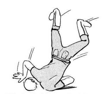
Young males in the age group of 15 to 25 years are most commonly affected. 70% of traumatic brachial plexus injuries occur secondary to motor vehicle accidents and, of these, 70% involve motorcycles or bicycles.
A fall from a significant height or gunshot wound may also result in brachial plexus injury.
Mechanism of Traumatic Brachial Plexus Injury
Mostly the trauma is closed and nerve injury occurs from traction [95%] and compression. The nerves may rupture, avulse from the root or be intact but significantly stretched.
The nerve can be injured at the root, the anterior branches of the spinal nerves, the trunk, the cord, and the peripheral nerve.
[Also read anatomy of brachial plexus]
Root injuries may postganglionic (or infraganglionic) or preganglionic (or supraganglionic) depending on whether lesions are distal to the dorsal root ganglion or proximal.
[Preganglionic injuries cause avulsions from the spinal cord resulting in separation of motor nerve fibers from the motor cell bodies in the anterior horn cells of spinal cord. However, The sensory fibers and cell bodies are still connected at the dorsal root ganglion but fibers entering the dorsal spinal column have been disrupted. Thus, sensory nerve action potentials are preserved in patients with supraganglionic injuries but motor are not. There is disruption of both motor and sensory pathways in postganglionic injuries leading to abnormalities of both motor action potentials and sensory nerve action potential.]Root avulsions can occur when the traction force overcomes the fibrous support around the roots [peripheral avulsion injury] or there is a severe bending of the spinal cord due to cervical injury [central avulsion injury]. Former are much more common.

Supraclavicular regions of brachial plexus are injured more commonly as compared to the infraclavicular region. The roots and trunks are more commonly injured in comparison to the divisions, cords, or terminal branches. More than one level of brachial plexus can be involved.
Upper brachial plexus injuries occur when the head and neck are violently moved away from the ipsilateral shoulder leading to stretch, avulsion, or rupture of the upper roots (C5, C6, C7). Lower roots are preserved.
Lower elements of the plexus (C8, T1) can be injured in forceful abduction and traction of the shoulder. Distal infraclavicular lesions can be associated with axillary arterial rupture.
70 to 75% of injuries are found in the supraclavicular region. The remaining plexus injuries are infraclavicular.
Open injuries are less common and result in transection. Iatrogenic injuries have been reported.
Presentation of traumatic Brachial Plexus Injury
The patient may present with pain in the neck and the shoulder, paresthesias, weakness or heaviness in the limb. Decreased pulses may be a sign of vascular injury.
In patients with significant trauma, the patient needs to be stabilized and resuscitated and diagnosis may be delayed. Suspicion of brachial plexus injury should be maintained in a patient with significant shoulder girdle injury, first rib injuries, or axillary artery injuries.
Dramatic shoulder swelling and decreased or absent pulses suggest vascular injury. Clavicle fractures often are palpable. Concomitant injuries should be noted.
Examination of brachial plexus and branches requires an awake and cooperative patient.
Testing muscles function and sensations supplied by different branches would indicate the site of the lesion. A brief summary is given below. Anatomical variations should be kept into mind.
- C5 – Shoulder abduction, extension, and external rotation, and some elbow flexion.
- C6 – Elbow flexion, forearm pronation, and supination, some wrist extension.
- C7 – Diffuse loss of function in the extremity without complete paralysis of a specific muscle group, elbow extension
- C8 – Finger extensors, finger flexors, wrist flexors, hand intrinsics
- T1 – Hand intrinsics
An injury to the long thoracic nerve or dorsal scapular nerve suggests a higher (more proximal) level of injury [both nerves originate from roots].
Presence of Horner’s syndrome indicates interruption of the sympathetic ganglion and avulsion of the T1 root.
An active and passive range of motion and reflexes are part of neurological examinations.
The spine should be essentially examined for ruling out/confirming the concomitant spinal injury.
A vascular examination to rule out the arterial injury is part of the examination
Laboratory Studies
Laboratory studies generally are not helpful for diagnosis, although they may be indicated in the routine evaluation of any trauma patient.
Imaging Studies
Radiographs
Standard radiographs should include cervical spine views, shoulder views (anteroposterior, axillary views), and a chest X-ray.
Following findings on x-rays should raise the concern for brachial plexus injury.
- An increase in distance between the spinous processes of the thoracic spine and the scapula suggesting scapulothoracic dissociation [Comparison may be done with opposite side]
- Cervical fractures
- Fractures of the transverse process fractures of the cervical vertebrae [ might indicate root avulsion at the same level]
- Fracture of the clavicle
- Fractures of first, second rib
- Elevated hemidiaphragm [indicates phrenic nerve is injured]
Arteriography
Arteriography may be indicated in cases where the vascular injury is suspected.
CT
CT scan of the neck can be done as a part of the evaluation. CT chest may reveal subclavian vessel injuries, scapular fractures, humeral fractures, and thoracic spine fractures.
Myelography/CT Myelography
Myelography should be delayed for at least 4 weeks so that meningocele at the affected level is allowed to form. CT myelography can detect lower concentration of the contrast as compared to plain myelography but is prone to greater artifacts.
MRI
MRI is helpful in evaluating the patient with a suspected nerve root avulsion. It is noninvasive and is can visualize the plexus whereas CT/myelography shows only nerve root injury. MRI can also reveal large neuromas and inflammation or edema.
However, CT myelography remains the gold standard of radiographic evaluation for nerve root avulsion.
Sensory nerve action potentials
SNAPs are very helpful in differentiating preganglionic from postganglionic injuries. A sensory nerve action potential confirms a preganglionic lesion.
Electromyography
If no signs of denervation are apparent in a paralyzed muscle by 3 weeks after injury, EMG can be used to confirm neuropraxia.
Somatosensory Evoked Potential
The presence of SSEPs suggests continuity between the peripheral nervous system and the CNS via the dorsal root ganglion.
Treatment of Brachial Plexus Injury
Brachial plexus injury was treated by the conservative method in the past which consisted of monitoring for 12-18 months. After that, any residual deficit was considered permanent. Shoulder fusion, elbow fusion, wrist and finger tenodesis and transhumeral amputation as required were done.
However, with improvement in treatment, surgical options are now available which are primary repair, soft-tissue reconstruction, nerve grafting and nerve transfers (neurotization).
Clear consensus regarding surgical timing and surgical indications is lacking. Surgery is not performed in presence of joint contractures, edema, advanced age of the patient and uncooperative patient.
Immediate exploration with possible end-to-end repair may be indicated in some cases of open injury caused by a sharp object. If the surgery is delayed, the patient is put on physical therapy, as in blunt trauma and avulsion injury, delay of 3-4 weeks.
In case of stretch injuries, spontaneous recovery should be watched for some time.
Reconstruction procedures may need to be done in a staged manner.
Neurotization or nerve transfers can be performed to accelerate recovery from preganglionic injuries using spinal accessory nerve, intercostal nerves, and the medial pectoral nerve, usually within 6 months of the trauma.
External neurolysis should be performed for nerves in continuity that exhibit a nerve action potential.
As noted before postganglionic lesions do not have sensory nerve action potentials.
Physical therapy and bracing often are used over the prolonged postoperative period to prevent contractures, to keep joints supple after surgery.
The prognosis is highly variable. It depends not only on the nature of the injury but also on the age of the patient and the type of procedure performed.