• Skip to primary navigation
  • Skip to main content
  • Skip to primary sidebar
  • Home
  • Online Consultation
  • About
  • Newsletter/Updates
  • Contact Us
  • Policies

Bone and Spine

Orthopedic health, conditions and treatment

  • General Ortho
  • Procedures
  • Spine
  • Upper Limb
  • Lower Limb
  • Pain
  • Trauma
  • Tumors

Bursae Around Shoulder

By Dr Arun Pal Singh

In this article
    • Different Bursae Around Shoulder
      • Subacromial bursa or Subacromial-subdeltoid Bursa
      • Subscapular Bursa
      • Subcoracoid bursa
      • Supra-acromial Bursa
      • Coracoclavicular  Bursa
      • Related

There are 5-6 bursae around the shoulder. Bursae are the synovial fluid-filled sacs that are present between two surfaces that rub on motion. The purpose of the bursa is to reduce friction and protect the moving tissue.

Often these are found at tendon-tendon and tendon-bone interfaces.

There are five main bursae around shoulder. They include:

  • Subacromial-subdeltoid bursa
  • Subscapular recess
  • Subcoracoid bursa
  • Coracoclavicular bursa
  • Supra-acromial bursa

Sometimes authors include medial extension of  subacromial-subdeltoid bursa (SASD) as sixth bursal space.

The bursae have free nerve endings for pain perception and mechanoreceptors for providing proprioceptive information of shoulder joint position.

Thus bursae might have roles beyond lubrication and cushion as well. Different bursae around the shoulder are discussed below.

bursae arund shoulder
Figure 1: Diagram of normal bursae surrounding the shoulder joint: (1) subacromial-subdeltoid bursa, (2) subscapular recess, (3) subcoracoid bursa, (4) coracoclavicular bursa, (5) supra-acromial bursa and (6) medial extension of subacromial-subdeltoid bursa.
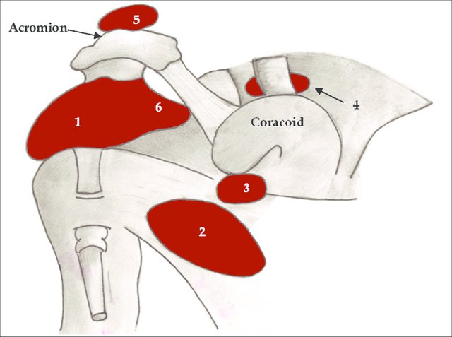
Image Credit: ResearchGate

Different Bursae Around Shoulder

Subacromial bursa or Subacromial-subdeltoid Bursa

The subacromial-subdeltoid (SASD) bursa comprise of two bursae that lie between the rotator cuff tendons and the undersurface of the acromion. They are located deep in the deltoid muscle and acromioclavicular joint but overlie the bicipital groove and rotator cuff.

The coracoacromial ligament, acromion bone, and the deltoid form the superior extent whereas the humeral head, the shoulder joint, and the supraspinatus form the inferior extent.

The main function of the bursa is to reduce friction in the space under the acromion.

Some authors keep subacromial and subdeltoid bursae as separate entities.

SASD bursitis is a common cause of shoulder pain and can occur due to many causes including:

  • Shoulder impingement
  • Tears of rotator cuff
  • Infection
  • Reaction to the glenohumeral joint disease such as crystal arthropathy

Pain between 60 and 120 degrees of abduction is considered indicative of subacromial deltoid bursitis.

Fluid-filled, SASD bursa is visible both on ultrasound and MRI but x-ray may be normal. However, MRI is rarely needed for diagnosis of bursitis.

Rest, anti-inflammatory drugs are the mainstay of the treatment. In selected cases, intrabursal steroid injections can be given. Surgical removal of the bursa is rarely needed.

Read more about Subacromial Bursitis

Subscapular Bursa

The subscapular bursa is also called subscapularis recess and it is a normal extension of the glenohumeral joint capsule in between the superior and middle glenohumeral ligaments. The subscapularis is a muscle of the shoulder region.

The bursa lies between the subscapularis muscle and the chest wall. Subscapular bursitis results in pain and popping sensation.

Pain and a popping sensation are typical complaints. Working overhead, reaching up and forward is painful.

A swollen bursa is visible on MRI.

Subcoracoid bursa

This bursa lies between the coracoid process and combined tendons of the short head of the biceps and coracobrachialis. It is superior to the subscapularis tendon.

This bursa decreases the friction between the subscapularis tendon and the tendons of the short head of biceps and coracobrachialis during the rotation of the humeral head.

Thus it facilitates internal and external rotation of the shoulder.

The subcoracoid bursa does not communicate with the glenohumeral joint. However, it may have communications with the subacromial bursa.

In subcoracoid bursitis, the passive external rotation in 90 degrees of abduction is negative and the passive external rotation in 0 degrees is painful.

A contrast fluid injected into the glenohumeral joint, thus, normally will not show in subcoracoid bursa during arthrography. But if it shows, it implies full-thickness rotator cuff tear of the shoulder.

Supra-acromial Bursa

As the name suggests, this bursa is located on the superior aspect of the acromion. It also does not communicate with the glenohumeral joint.

There are very few reports of supracromial bursitis in the literature.

Coracoclavicular  Bursa

The coracoclavicular ligament is composed of two parts, the conoid and trapezoid ligaments. These ligaments are continuous inferiorly at the coracoid process attachment but separate at an angle before they attach to the inferior aspect of the clavicle superiorly.

A bursa separates the trapezoid ligament and the conoid ligament. There could be up to three coracoclavicular ligament bursae. These bursae can have bursitis though it is not that common.

Related

Spread the Knowledge
2
Shares
 
2
Shares
2    

Filed Under: Hand and Upper Limb

About Dr Arun Pal Singh

Arun Pal Singh is an orthopedic and trauma surgeon, founder and chief editor of this website. He works in Kanwar Bone and Spine Clinic, Dasuya, Hoshiarpur, Punjab.

This website is an effort to educate and support people and medical personnel on orthopedic issues and musculoskeletal health.

You can follow him on Facebook, Linkedin and Twitter

Primary Sidebar

Browse Articles

repetitive strain injury

Repetitive Strain Injury – Causes, Symptoms and Treatment

Repetitive strain injury or RSI is a term describing the conditions where symptoms develop due to build of damage over the period, owing to repetitive motion. To say some person has repetitive strain injury or repetitive stress injury, other pathologies should not be present and the pain complaints can be related to repeated activities over […]

Different Bone Cells - Origin and Fate

Bone Cells – Types and Function

A cell is a basic building unit of any tissue or organ. Bone cells constitute bone tissue which are arranged to form the structure we see outwardly. Following types of bone cells are found in the bone. Osteoclasts Osteoblasts Osteocytes Lining cells Different types of bone cells are programmed for kinds of functions. Osteoclasts The […]

Muscles of Hip and Thigh

Muscles of Hip

Muscles of the hip joint are those muscles that cause movement in the hip. The muscles of the hip consist of four main groups. Gluteal Group The gluteal muscles cover the lateral surface of the ilium and include Gluteus maximus Gluteus medius Gluteus minimus Tensor fasciae latae Adductor group This group includes Adductor brevis Adductor […]

Woven Bone Matrix

Woven Bone and Lamellar Bone

The major constituent of the is the bone matrix. Woven and lamellar bone are the terms based on the microscopic differentiation of the bone. Bone is formed by the hardening of this matrix entrapping the cells. For example, when are trapped in the matrix, they become osteocytes. The inorganic is mainly crystalline mineral salts and […]

Hydatid Cyst of Bone

Hydatid Disease of Bone

Hydatid disease of bone is very rare and is often misdiagnosed because of non-specific presentation and radiology. As its management is quite different from other conditions involving the bone, it is important to consider the differential of Hydatid disease of the bone in lucent bone lesions, especially where the echinococcosis is prevalent. Hydatid Disease or […]

Anatomy of Brachial Plexus

Brachial Plexus Anatomy

The brachial plexus is a network of nerves formed by the anterior rami of the lower four cervical nerves and first thoracic nerve roots and proceeds through the neck, the axilla, and into the arm. The nerves coming out from this supply upper limb and shoulder girdle. Structure of Brachial Plexus The brachial plexus consists […]

Plantar Fascia Anatomy

Plantar Fasciitis – Causes and Treatment

Plantar fasciitis is a common cause of heel pain and occurs due to degenerative changes at the insertion of the plantar fascia on calcaneal tuberosity. The plantar fascia is the most common cause of heel pain for which professional help is sought. A heel spur is commonly found in plantar fasciitis patients, but many asymptomatic […]

© Copyright: BoneAndSpine.com
Manage Cookie Consent
The site uses cookies. Please accept cookies for a better visiting experience.
Functional Always active
The technical storage or access is strictly necessary for the legitimate purpose of enabling the use of a specific service explicitly requested by the subscriber or user, or for the sole purpose of carrying out the transmission of a communication over an electronic communications network.
Preferences
The technical storage or access is necessary for the legitimate purpose of storing preferences that are not requested by the subscriber or user.
Statistics
The technical storage or access that is used exclusively for statistical purposes. The technical storage or access that is used exclusively for anonymous statistical purposes. Without a subpoena, voluntary compliance on the part of your Internet Service Provider, or additional records from a third party, information stored or retrieved for this purpose alone cannot usually be used to identify you.
Marketing
The technical storage or access is required to create user profiles to send advertising, or to track the user on a website or across several websites for similar marketing purposes.
Manage options Manage services Manage vendors Read more about these purposes
View preferences
{title} {title} {title}
 

Loading Comments...