• Skip to primary navigation
  • Skip to main content
  • Skip to primary sidebar
  • Home
  • TeleConsult
  • About
  • Newsletter/Updates
  • Contact Us
  • Policies

Bone and Spine

Orthopedic health, conditions and treatment

  • General Ortho
  • Procedures
  • Spine
  • Upper Limb
  • Lower Limb
  • Pain
  • Trauma
  • Tumors

Cast Syndrome or Superior Mesenteric Artery syndrome

By Dr Arun Pal Singh

In this article
    • Cause of Cast Syndrome
    • Relevant Anatomy
    • Clinical Presentation of Cast Syndrome
    • Imaging
      • X-rays
      • CT
    • Lab Tests
    • Treatment of Cast Syndrome
    • Prevention
      • Related

Cast syndrome is also known as superior mesenteric artery syndrome. It is an uncommon complication in the treatment of orthopedic conditions. It results from obstruction of the third portion of the duodenum due to the narrowing of the angle between the abdominal aorta and superior mesenteric artery.

Other names for cast syndrome are

  • Wilkie’s syndrome
  • Duodenal ileus

This was first described by Von Rokitansky in 1842 and later by Wilkie.

It is a rare syndrome and is more common in young females.

Mortality is around 33%.

It should be kept in mind that this obstruction can occur in absence of plaster also because there are many causes to mesenteric artery obstruction.

Cause of Cast Syndrome

scoliosis-jacketsMost cases involve young adults.

More than half of these cases have patients with scoliosis or kyphosis or treatment of hip conditions.

It has been seen after casting with

  • Body jackets
  • Shoulder spicas
  • Hip spicas

Thus the common denominator is extensive coverage of the abdomen and chest.

Relevant Anatomy

The problem usually is located at the junction of the third and fourth parts of the duodenum, where the duodenum is bound by the ligament of Treitz. The duodenum passes across the anterior aspect of the lumbar spine from right to left at the level of the first and second lumbar vertebrae. Just above this point, the superior mesenteric artery arises from the abdominal aorta and passes downward with its accompanying veins in the mesentery.

compression of duodenum in cast syndrome
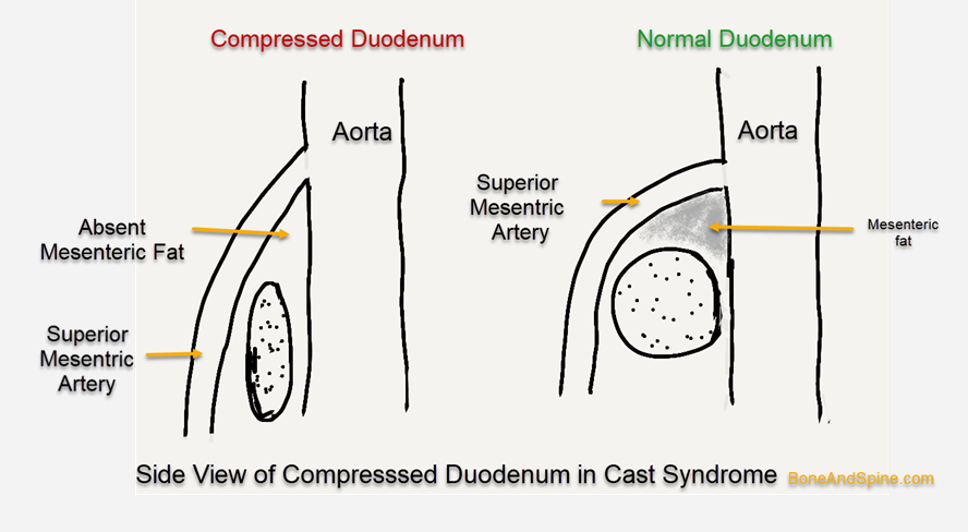
There is the potential for compression of the third portion of the duodenum between lumbar spine and aorta posteriorly and the mesentery and vessels anteriorly.

Two contributing factors are

  • Recumbency or lying down- causes the weight of the mesentery to lie against the duodenum
  • Increased lumbar lordosis, which tends to displace the duodenum anteriorly.

Gastric distention may aggravate the situation by forcing the remainder of the abdominal contents more distally and stretching the mesenteric vessels further leading to a spectrum of symptoms.

Clinical Presentation of Cast Syndrome

The signs and symptoms of cast syndrome are typical of upper intestinal obstruction.

They may develop insidiously or after several weeks after cast application or surgery.

The initial symptom is a feeling of fullness followed by nausea and vomiting.

Abdominal distention is obscured in the presence of a body jacket.

Following is the usual sequence of symptoms and signs

  • Abdominal Distension
  •  Vomiting
    • Intermittent in the early stages
    • Becomes pernicious later
  • Dehydration and metabolic alkalosis results
  • Progressive metabolic derangement occurs
  • Decrease in the passage of urine – oliguria, and shock may occur.

When these signs and symptoms occur in a patient who is in a body cast or who has had spinal trauma (including surgery), the diagnosis should be suspected.

Imaging

X-rays

X-rays of the abdomen may show early gastric dilatation.  Contrast may help to reveal the distention of the stomach and the proximal portion of the duodenum. Usually, there is a  sharp cutoff pattern at the region where the arteriomesenteric pedicle crosses the duodenum.

X-rays help to differentiate this syndrome from cholelithiasis, pancreatitis, gastric or duodenal ulcer, and high intestinal obstruction.

CT

CT abdomen with IV contrast helps to reveal the following

  • Dilatation of stomach and proximal duodenum
  • Decompression of rest of the duodenum and small bowel
  • Aortomesenteric distance [normal 10-28 mm]
    • Can be as low as 2 mm in cast syndrome
  • Aortomesenteric angle
    • Normal – 25–60 degrees
    • Can be as low as 7 degrees in cast syndrome

Lab Tests

Most of the biochemistry blood works would come normal. In later stages electrolyte imbalance may occur.

Treatment of Cast Syndrome

  • Removal of the offending plaster if any
  • The absolute dietary restriction – Nothing by mouth
  • Nasogastric tubing to conduct suction of the gastric secretions
  • Intravenous fluids are essential to maintain hydration
  • Correct any electrolyte or acid-base abnormalities
  • Positioning the patient on the left side or prone relieves some of the effects of gravity in pulling the mesenteric pedicle against the duodenum. With the patient prone, tipping the bed to tilt the head down further decreases pressure from the pedicle.
  • If these conservative measures are not sufficient to reverse the process, the treatment of choice is surgery duodenojejunostomy or gastrojejunostomy.

Prevention

Cast syndrome may be prevented by avoiding constrictive body casts that accentuate lumbar lordosis or prevent normal changes in abdominal contour. Patients at risk should be turned frequently or encouraged to move from side to side.

Related

Spread the Knowledge


Filed Under: Spine

About Dr Arun Pal Singh

Arun Pal Singh is an orthopedic and trauma surgeon, founder and chief editor of this website. He works in Kanwar Bone and Spine Clinic, Dasuya, Hoshiarpur, Punjab.

This website is an effort to educate and support people and medical personnel on orthopedic issues and musculoskeletal health.

You can follow him on Facebook, Linkedin and Twitter

Primary Sidebar

Browse Articles

mri-c5-c6-dislocation

Subaxial Cervical Spine Injury Classification System or SLICS

The Subaxial Cervical Spine Injury Classification (SLIC) system is a classification system for subaxial cervical spine injury. This is one of the recommended classification systems for lower cervical injury. Not only it helps to determine the injury pattern and its severity but also it guides the treatment considerations and prognosis. The classification is similar to TLICS […]

Wound debridment becomes necessary in such cases

Wound Debridement Types and Methods

Wound debridement or debridement is the process of removing infected, damaged, dead, nonviable tissue or foreign matter wounds so as to improve the healing potential of the remaining healthy tissue. Wound debridement can be achieved with surgery or by nonsurgical means. Why Wound Debridement is Done? A wound that contains tissue which is dead or […]

Xray of Madelung Deformity

Madelung Deformity Causes and Treatment

Madelung deformity is an abnormality of the wrist caused by a growth disturbance volar-ulnar distal radial physis that causes Partial deficiency of growth of distal radial physis Excessive radial inclination and volar tilt Ulnar carpal impaction The tilt in the distal radius is caused by inequality of growth. This leads to volar or palmar translation […]

Medial malleolus fracture

A Case of Medial Malleolus Fracture Fixed By Percutaneous Fixation

Medial malleolus fracture is a fracture of the distal medial projection of tibia bone called medial malleolus. Medial malleolus and lateral malleolus contribute forms the socket of the ankle joint where talus articulates. There are three malleoli in the ankle. The medial and the posterior arise from the tibia. A counterpart called lateral malleolus is […]

Gluteal intramuscular site injection

Gluteal Region Anatomy and Significance

The gluteal region is an anatomical area located posteriorly to the pelvic girdle, at the proximal end of the femur. In approximate terms, it can be called the area of buttocks. There are two gluteal regions, left and right. The muscles in this region move the lower limb at the hip-joint. Cutaneous Nerves, Vessels and […]

Xray of Charcot Joint of Foot

Charcot Joint Disease

Charcot joint or Charcot arthropathy is the name given to neuropathic joint disease is a condition that causes progressive destruction of bone and soft tissues of joints especially weight-bearing joints and may also cause disruption of the bony architecture. Charcot joint disease can occur at any joint but most commonly in the lower extremity, at […]

rib-cage

Anatomy of Thoracic Cage or Rib Cage

The thoracic cage or rib cage is the skeletal framework that encloses the contents of the thorax. The thoracic cage or rib cage along with its contents form the thorax portion of the body. Roughly speaking, this is the area of the chest. The Thoracic cage consists of the 12 pairs of ribs with their […]

© Copyright: BoneAndSpine.com
Manage Cookie Consent
The site uses cookies. Please accept cookies for a better visiting experience.
Functional Always active
The technical storage or access is strictly necessary for the legitimate purpose of enabling the use of a specific service explicitly requested by the subscriber or user, or for the sole purpose of carrying out the transmission of a communication over an electronic communications network.
Preferences
The technical storage or access is necessary for the legitimate purpose of storing preferences that are not requested by the subscriber or user.
Statistics
The technical storage or access that is used exclusively for statistical purposes. The technical storage or access that is used exclusively for anonymous statistical purposes. Without a subpoena, voluntary compliance on the part of your Internet Service Provider, or additional records from a third party, information stored or retrieved for this purpose alone cannot usually be used to identify you.
Marketing
The technical storage or access is required to create user profiles to send advertising, or to track the user on a website or across several websites for similar marketing purposes.
Manage options Manage services Manage vendors Read more about these purposes
View preferences
{title} {title} {title}
 

Loading Comments...