Last Updated on October 29, 2023
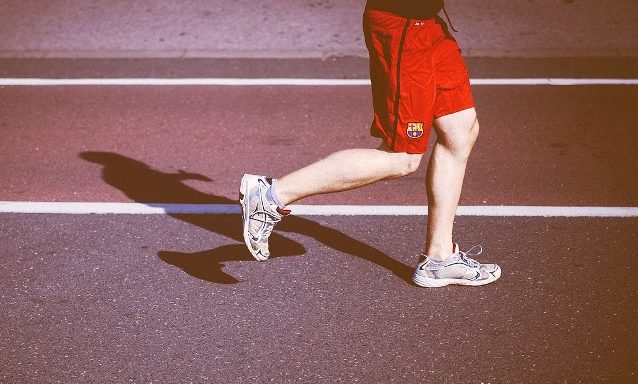
Chronic exertional compartment syndrome is a condition characterized by exercise-induced pain that is relieved by rest. It occurs from repetitive loading or exertional activities in athletes, long-distance runners, basketball players, skiers, and soccer players.
It is most commonly seen in legs but can involve any compartment of the extremities. It is the second most common exercise-induced leg syndrome. It is often seen in the third decade of life and found in runners or those sportspersons who run a lot for their sport.
Leg, thigh, and forearms are the most common sites involved in chronic exertional compartment syndrome.
Forearms are typically seen involved in racers motocross racers.
Males and females are affected equally though some studies report a higher incidence in males.
Pathophysiology of Chronic Exertional Compartment Syndrome
The pathology of chronic exertional compartment syndrome is not well established.
It is thought that the local muscle metabolism cannot go fast enough to clear the metabolic waste products.
It is suggested that affected persons may have a lower density of capillaries compared to asymptomatic individuals.
Fascial defects have been found to be associated with chronic exertional compartment syndrome. 40% of people with exertional compartment syndrome have these fascial defects but only 5% of asymptomatic people have such defects
In chronic exertional compartment syndrome, however, the pressure between successive contractions remains high and impedes blood flow. As the pressure rises, arterial flow during muscle relaxation decreases, and the patient gets muscle cramping.
In a normal person, raised compartmental pressures to normalize within 5 minutes after cessation of exercise but in patients with the syndrome, the pressures may remain elevated for 30 minutes or longer.
When tissue blood flow is diminished further, muscle ischemia and subsequent cell edema worsen. This vicious cycle of worsening tissue perfusion continues to propagate.
With further destruction, myoglobin is released into the circulation which may result in renal injury.
Various mechanisms have been suggested as to the cause of this tissue ischemia
- Arterial spasm
- Capillary obstruction
- Venous outflow obstruction.
- Muscle hypertrophy and/or fascial inflexibility
- Exercise resulting in myofibril damage and release of protein-bound ions.
No single theory has been overwhelmingly accepted.
Clinical Presentation of Chronic Exertional Compartment Syndrome
Patients report pain or tightness, cramping, burning, or aching over the affected compartment during exercise. The affected extremity may feel weak.
The pain may develop predictably at a specific point in an exercise session leading to disruption of activity. Some runners may be able to continue running with a modified flatfoot strike.
The patient may also complain of weakness of muscles. Paresthesia or dysesthesia may develop in the distribution of the affected nerve.
Symptoms tend to subside with rest. Any persistent symptoms are usually minimal during normal daily activities.
The patient may note bumps or herniations over the affected compartment. The patient usually denies any edema, temperature changes, or color changes of the affected extremity.
Physical examination is usually normal unless the patient has recently exercised. The calf may be firmer or tense on examination.
Depending on the involved compartment in the leg, the patient may complain of following weaknesses.
- Anterior compartment
- Weakness on dorsiflexion
- Loss of sensation in the web of the first toe
- Involvement of the deep peroneal nerve.
- Lateral compartment
- Weakness upon inversion
- Loss of sensation on the anterolateral part of the shin and the dorsum of the foot
- Involvement of the superficial peroneal nerve.
- Deep posterior compartment
- Weakness in the foot muscles
- Loss of sensation in the foot arch
- Due to the involvement of the tibial nerve.
The distal neurovascular examination is normal. If not, consider other pathologies.
These patients usually do not have tenderness over the posterior medial tibial cortex in the distal leg. This contrasts with medial tibial stress syndrome, in which tenderness is typically located in this area.
There is no focal tenderness. Patients with chronic exertional compartment syndrome usually do not present with focal tenderness with overlying edema. This finding is more indicative of a stress fracture.
Differential Diagnoses
- Deep Venous Thrombosis
- Diphyllobothriasis [a parasitic infection]
- Hypothyroid Myopathy
- Lumbosacral Radiculopathy
- Myopathies
- Nerve Entrapment Syndromes
- Spinal Stenosis
- Tumor
Diagnosis
The diagnosis of chronic exertional compartment syndrome is often based on the clinical history, the physical examination findings, and the exclusion of various differential diagnoses.
Compartment pressure readings with and without exercise are the gold standard for the diagnosis.
Lab Studies
Lab studies are generally not diagnostic. Specific tests to rule out other causes of lower leg pain can be done. These include
- Complete blood cell count
- Complete metabolic panel
- Thyroid function tests
- Erythrocyte sedimentation rate
- Serum creatine kinase and myoglobin level
- Identifies myopathy or rhabdomyolysis
- Urine myoglobin
- Rhabdomyolysis
- D-dimer level
- Deep venous thrombosis
Imaging
Imaging studies are not helpful in the diagnosis but may rule out related disorders
Anteroposterior, lateral, and oblique view radiographs are done. X-rays of the spine may be done where spine pathology is suspected.
Bone scanning helps exclude stress fracture, periostitis, and malignancy of the lower extremity.
CT/MRI can help rule other significant causes of chronic lower leg pain.
Compartment Pressure Testing
Compartment pressure readings with and without exercise are the gold standard for the diagnosis. Pain reproduced during exercise in combination with elevated compartment pressures can confirm the diagnosis. If symptoms are not reproduced with exertion, the diagnosis is less certain.
For this procedure, a large-bore needle or a wick catheter is inserted into the affected muscular compartment and is then connected to a solid-state pressure monitor.
Pedowitz et al criteria
- A pre-exercise/rest pressure of 15 mm Hg or higher
- A 1-minute postexercise pressure of 30 mm Hg or higher
- A 5-minute postexercise pressure of 20 mm Hg or higher
The diagnosis can is made if just 1 of the above criteria is met. But more than one points increase the confidence level of the diagnosis.
Treatment of Chronic Exertional Compartment Syndrome
Non-operative Treatment
Nonoperative treatment involves a decreased loading of the affected compartment by decreasing activity.
The activity level gradually is increased then. Aquatic exercises, massage, and stretching exercises are also recommended.
Physical therapy includes reduction of activity, cross-training exercises and muscle stretching before initiating exercise.
Symptoms generally recur when the patient returns to exercise.
If non-operative therapy is not successful, the patient should be considered for fasciotomy.
Early mobilization, as soon as is feasible, is recommended after the surgery to minimize the adhesions.
Activity can be upgraded to stationary cycling and swimming after healing of the surgical wounds.
Running is integrated into the activity program at 3-6 weeks. Full activity is introduced at approximately 6-12 weeks, with a focus on speed and agility.
Prognosis
Surgery produces good results and ability to return to sports. For unknown reasons, the deep posterior compartment does not respond as quickly or as well
Fasciotomy of the anterior compartment has a better outcome than fasciotomy of the posterior compartment. [
Furthermore, the rehabilitation phase is longer for patients with deep posterior compartment fasciotomy than it is for those who undergo anterior compartment fasciotomy. The reasons for this difference in outcome remain unclear.
References
- Claes T, Van der Beek D, Claes S, Verfaillie S, Bataille F. Chronic exertional compartment syndrome of the forearm in motocross racers. Presented at: The European Sports Medicine Congress; Hasselt, Belgium; May 14-16, 2003.
- Blackman PG. A review of chronic exertional compartment syndrome in the lower leg. Med Sci Sports Exerc. 2000 Mar. 32(3 suppl): S4-10.
- Roscoe D, Roberts AJ, Hulse D. Intramuscular compartment pressure measurement in chronic exertional compartment syndrome: new and improved diagnostic criteria. Am J Sports Med. 2015 Feb. 43 (2):392-8.
- Clayton JM, Hayes AC, Barnes RW. Tissue pressure and perfusion in the compartment syndrome. J Surg Res. 1977 Apr. 22(4):333-9
- Amendola A, Rorabeck CH, Vellett D, et al. The use of magnetic resonance imaging in exertional compartment syndromes. Am J Sports Med. 1990 Jan-Feb. 18(1):29-34
- Styf JR, Körner LM. Chronic anterior-compartment syndrome of the leg. Results of treatment by fasciotomy. J Bone Joint Surg Am. 1986 Dec. 68(9):1338-47.
- Hutchinson MR, Ireland ML. Common compartment syndromes in athletes. Treatment and rehabilitation. Sports Med. 1994 Mar. 17(3):200-8