• Skip to main content
  • Skip to primary sidebar
  • Skip to footer
  • General Ortho
  • Procedures
  • Spine
  • Upper Limb
  • Lower Limb
  • Pain
  • Trauma
  • Tumors
  • Newsletter/Updates
  • About Us
  • Contact Us

Bone and Spine

Orthopedic health, conditions and treatment

Complex Regional Pain Syndrome or CRPS

By Dr Arun Pal Singh

In this article
    • Causes of CRPS
    • Pathophysiology
    • Revised CRPS Clinical Diagnostic Criteria
    • Clinical Features
    • Differential Diagnoses
    • Lab Studies
    • Imaging
    • Other Tests
    • Treatment of CRPS
      • Drugs
      • Interventional Procedures
      • Physical Therapy
      • Surgical Care
    • Prognosis
    • References

Complex regional pain syndrome or CRPS is a chronic painful condition causing intense pain, usually in the arms, hands, legs, or feet. The cause of condition is poorly understood and there is no cure. The management involves control of symptoms by various modalities.

The condition It may happen after an injury to a nerve or other tissues.

In th past, it was known by many other names like reflex sympathetic dystrophy, Sudeck’s atrophy, reflex neurovascular dystrophy or algoneurodystrophy.

Causalgia was the name given to a condition associated with nerve injury.

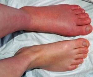
The condition involves dramatic changes in skin temperature, color, or texture, intense burning pain, swelling, extreme skin sensitivity and dysfunction of the affected body part.

Depending on the presence or absence of nerve involvement it can be on of the two types.

  • Type I [Reflex sympathetic dystrophy] – No demonstrable nerve lesions.
    • Symptoms < 2 months- acute
    •  >2 months – chronic.
  • Type II [causalgia]- Associated with nerve damage.

The incidence of CRPS type I is higher than that of CRPS type II.

CRPS affects all races and is more common in females. The ratio varies from 2:1 to 4:1.

Its peak incidence between 37 and 50 years.

CRPS is known to affect all age groups but occurrence in children is extremely rare.

Most often, CRPS involves extremities but locations like genitalia or the nose have been reported. The single limb is involved in most of the cases. Bilateral involvement is seen in about 5% cases. Rarely more than two limbs could be involved.

complex-regional pain syndrome lower limb
Image Credit: Orthobullets

Causes of CRPS

As noted before, type II is associated with nerve injury.

Trauma to the limb is the most common cause of type I CRPS accounting for more than 90% of patients with CRPS type I.

Associated injuries are or injuries precipitating are

  • Sprain/strain
  • Surgical wounds
  • Fractures
  • Crush injury/contusion
  • Rarely in following injuries
    • Venous puncture
    • Lacerations
    • Burns
    • inflammatory Conditions
    • Electric shock
  • Cases with unknown causes occur in 5% [probably forgotten minor injuries]
  •  Visceral lesions or  CNS lesions [controversial]

Pathophysiology

The exact cause of the condition is not known.

It is believed persistent painful stimuli from an injured region causes peripheral and central sensitization.

Pain fibers have abnormally heightened sensation leading to pain and hyperalgesia.

There is also a central somatosensory misinterpretation of the nonpainful mechanical stimuli like a light touch. Plus there is an impairment of CNS processing leads to motor effects such as weakness or tremor in the affected area.

The disturbances within the sympathetic nervous system lead to sympathetic hyperactivity which affects the injured area adversely.

Also suggested is an augmented inflammatory response coupled with impaired healing. This makes the CRPS refractory.

Decreased use of an injured body by casting or splinting has been postulated as a cause in some patients with CRPS.

But all these are part of the hypothesis only.

Revised CRPS Clinical Diagnostic Criteria

[International Association for the Study of Pain]/

A clinical diagnosis of complex regional pain syndrome can be made when the following criteria are met:

  • A continuous pain which is disproportionate to any inciting event
  • At least 1 symptom reported in at least 3 of the following categories
    • Sensory
      • Hyperesthesia [Increased sensitivity]
      • Allodynia [pain due to non-painful stimuli due to central sensitization]
    •  Vasomotor[compared to healthy limb]
      • Temperature asymmetry
      • Skin color changes
      • skin color asymmetry
    •  Sudomotor/edema:
      • Edema
      • sweating
    • Motor/trophic
      • Decreased range of motion
      • Motor dysfunction ( weakness, tremor, dystonia etc)
      • Trophic changes in hair, nail, skin
  • At least 1 sign at the time of evaluation in at least 2 of the above categories
  • No other diagnosis explaining the signs and symptoms

It is important to note that there are no specific diagnostic tests and CRPS is diagnosed mainly clinically.

Clinical Features

The symptoms of CRPS I may begin immediately or even weeks after the initial injury.

Pain is the most common complaint. The pain is said to occur spontaneously and is not limited to region supplied by a single peripheral nerve. The pain is said to be burning, aching, throbbing, or tingling. The activity of the extremity worsens the pain.

The patient also complains of difficulty/inability to use the affected extremity.

The limb may feel as foreign [cognitive neglect] and required mental and visual attention to carry out the movement [motor neglect]. The limb also gets easily fatigued.

There is an asymmetry of the temperature of the skin when compared to the opposite limb.

The limb could be warmer [in two-thirds] or cooler than the opposite.

A physical examination may reveal

  • paresis, pseudoparalysis, or clumsiness, seen in 90% at some point in the disease
  • Limited range of motion
  • Tremors
  • Dystonia of affected hand or foot is seen in 10%
  • Muscle spasms [ 25% of patients]
  • Hypoesthesia [decreased sensation],  Hypothermethsia [decreased sense of temperature]
  • Loss of proprioception in some cases.
  • Anesthesia dolorosa – Loss of sensitivity to touch and severe pain is present.
  • Allodynia (ie, pain to touch), hyperpathia (exaggerated response to painful stimuli) [70-80%]
  • Abnormal sweating
  • Edema
  • Altered skin color [vasomotor changes]
  • Atrophy of the soft tissues, muscles, and bones [disuse also contributes]
  • Altered skin temperature [ 40-45% a warmer affected extremity, and 40-45% have a colder]
  • Decreased hair in the affected area
  • Altered nail growth

Differential Diagnoses

  • Neuropathy like diabetic polyneuropathy
  • Posttraumatic neuralgia
  • Plexopathy
  • Vascular disorders like  Raynaud phenomena
  • Psychological

Lab Studies

There is no specific confirmatory diagnostic test for the presence of CRPS.

But the role of investigations is to rule out other diagnostic considerations

  • Complete blood count
  • ESR
  • C-reactive protein
  • Antinuclear antibody
  • Rheumatoid factor
  • Complement fixation panel
  • Serum immunoelectrophoresis
  • Diabetes work up
  • Electromyography (EMG) and nerve conduction to determine nerve injury
  • Vascular studies of the affected limb(s)

Imaging

X-rays are suggestive of disuse and osteoporosis. These include Endosteal and intracortical excavation, subperiosteal resorption, demineralization of the region etc

3-phase bone scintigraphy is highly sensitive and specific in early CRPS that occurs after fracture and helps in differentiating it from other pain syndromes. The findings are

  • Characteristic homogeneous unilateral hypoperfusion [ perfusion phase, blood-pool phases]
  • Mineralization phase shows increased unilateral periarticular uptake. [highly sensitive and specific to CRPS

Other Tests

  • Quantitative sensory testing
  • Autonomic function testing
    • Infrared thermometry and thermography
    • Quantitative sudomotor axon reflex test
    • Thermoregulatory sweat test
    • Laser Doppler flowmetry.
  • Neurogenic inflammation markers [Interleukin-6, tumor necrosis factor-alpha, tryptase, and endothelin-1
    • Checked in fluid of artificially produced blisters
    • But may be present  evidence after the CRPS improves
  • Skin and muscle biopsies to look for atrophic changes
  • Peripheral nerve biopsies to look for any neural degeneration

Treatment of CRPS

Objective of the treatment is to restore functional extremity. The treatment is multidisciplinary where pain control plays a major role. Treatment is more effective when started early.

Following are the various treatment modalities.

Drugs

Pain Removal drugs

No clear-cut evidence for opioids or NSAIDs but Opioids still considered are part of treatment.

Tricyclic antidepressant

  • Amitriptyline
  • Desipramine

Gamma-aminobutyric acid (GABA) agonist

  • Intrathecally administered baclofen for dystonia
  • Gabapentin – mildly beneficial for pain and sensory symptoms

Calcium-regulating drug

  • Intranasal calcitonin administered significantly reduce pain
  • IV clodronate and alendronate
    • improve pain, swelling, and range of movement
    • mechanism of action unknown

Corticosteroids

  • Pulsed steroids can be considered if inflammatory signs and symptoms predominate.

Sodium channel blockers

  • IV lidocaine
  • Topical 5% lidocaine patch

Interventional Procedures

Sympathetic ganglion nerve blocks

Injections of local anesthetic around the sympathetic paravertebral ganglia that project to the affected part.

Intravenous regional sympathetic block

  • Regional IV applications of the agent to an isolated extremity blocked with a tourniquet
  • Various agents used are
    • Guanethidine – works by depleting norepinephrine, also has serotonergic and anticholinergic activity. 
    • Bretylium with or without lidocaine
    • Droperidol-  an alpha-adrenergic antagonist
    • Ketanserin, a serotonin type-2 antagonist

Intravenous phentolamine infusion

  • Alpha-1 adrenergic antagonist also has serotonergic, histaminergic, and cholinergic activities
  • The systemic activity allows for the simultaneous treatment of multiple body regions
  • Doses vary in different persons

Ketamine infusion

  • Blocks NMDA receptors
  • Objective outcome yet not validated.
  • Could be hospital-based or outpatient based.

Intravenous immunoglobulin

IV immunoglobulin reported providing pain relief. It also suggests the role of the immune system in pain of CRPS.

Epidural clonidine

Reported to be effective but associated with severe side effects.

Surgical sympathectomy

The procedure is not widely recommended as it has been found to have limited efficacy.

Spinal cord stimulation/neuromodulation

Spinal cord stimulation has been found to be an effective short-term treatment for CRPS and improved quality of life. 

Transcranial magnetic stimulation

It is a non-invasive procedure for the stimulation of the motor cortex but needs to be studied to explore the therapeutic role of magnetic stimulation in CRPS patients.

Physical Therapy

  • Most important component of treatment
  • Begin with the cautious mobilization of the part
  • Active isometric strengthening when resting pain subsides
  • Sensory desensitization programs
  • Patients who initially have less pain and better motor function are likely to benefit the most from physical therapy
  • Attentional training

Surgical Care

  • Surgical sympathectomy has been mentioned earlier
  • Surgical decompression in CRPS type II if there is a defined nerve [i.e. capral tunnel syndrome]
  • Amputation of the affected limb
    • Last resort
    • Rarely recommended

Prognosis

If the treatment is begun early good progress can be made. The prognosis is not always good in delayed cases. In extreme cases, amputation might be advised to get rid of the annoying and nonfunctional limb.

References

  • Rockett M. Diagnosis, mechanisms and treatment of complex regional pain syndrome. Curr Opin Anaesthesiol. 2014 Oct. 27(5):494-500.
  • Lee DH, Lee KJ, Cho KI, Noh EC, Jang JH, Kim YC, et al. Brain alterations and neurocognitive dysfunction in patients with complex regional pain syndrome. J Pain. 2015 Jun. 16 (6):580-6.
  • Drummond PD, Finch PM, Gibbins I. Innervation of hyperalgesic skin in patients with complex regional pain syndrome. Clin J Pain. 1996 Sep. 12(3):222-31.
  • Juottonen K, Gockel M, Silen T, Hurri H, Hari R, Forss N. Altered central sensorimotor processing in patients with complex regional pain syndrome. Pain. 2002 Aug. 98(3):315-23.
  • Veldman PH, Reynen HM, Arntz IE, Goris RJ. Signs and symptoms of reflex sympathetic dystrophy: prospective study of 829 patients. Lancet. 1993 Oct 23. 342(8878):1012-6.
  • Christensen K, Jensen EM, Noer I. The reflex dystrophy syndrome response to treatment with systemic corticosteroids. Acta Chir Scand. 1982. 148(8):653-5
  • Cepeda MS, Carr DB, Lau J. Local anesthetic sympathetic blockade for complex regional pain syndrome, Cochrane Database Syst Rev. 2005. 4:CD004598.
Spread the Knowledge
  • 14
    Shares
  •  
    14
    Shares
  • 1
  • 13
  •  
  •  
  •  

Filed Under: Pain Management

About Dr Arun Pal Singh

Arun Pal Singh is an orthopedic and trauma surgeon, founder and chief editor of this website. He works in Kanwar Bone and Spine Clinic, Dasuya, Hoshiarpur, Punjab.

This website is an effort to educate and support people and medical personnel on orthopedic issues and musculoskeletal health.

You can follow him on Facebook, Linkedin and Twitter

Reader Interactions

Comments

  1. Debbie Bishop says

    September 4, 2010 at 12:48 am

    I have had CRSD ror the past 16 months. And it is the most unbareable thing that I have ever had to go though in my life. I have had spinale blocks done 2 dirrerent times. Been in PT for 14 months. And I just had a knee replacement done about 13 weeks ago. In which has coused me to have a major major sat back. It is so hard for me to understand that there is NO answer or NO cure for it. Everyday I spend crying and in so much pain, at first my family did not understand the amount of PAIN that I was in, less alone why and h ow it worked. Sence I was told that I had CRSD I have ended up with High Blood Preasurer, Blood Suger Promblems, but most of all I am in so much pain all the time. And now there is some talk about having my leg taking off right above the knee. As for those of you that may not know or understand CRSD or RSD if you have anyone you know or love that does have CRSD, please do all the studing you can do to help the one you love. The one thing that I have learned is all emosnional streess will make the CRSD really bad. There is only one more thing left that I have not had done and that is the spine emplant and becouse of all the other medical prombles that I have there is no chance that I will have that done. I do know that I dont wont to have my leg cut of but if it will make the pain go away at lest some then I am really giving it some thought. Becouse I can not take it anymore. Would really like to hear from someone eles that has got the CRSD and hear what thier thought on this really really bad painful killer is. Thank you for taking your time to read my thoughts it means alot, maybe someone might be able to tell me of someone that would be able tell me of something eles that I maybe able to try or do. Thanks. May the good lord bless each and everyone of you. TAKE CARE.

Leave a Reply Cancel reply

Your email address will not be published. Required fields are marked *

This site uses Akismet to reduce spam. Learn how your comment data is processed.

Primary Sidebar

atlas vertebra

Surgery in Spinal Tuberculosis

Surgery in spinal tuberculosis is considered in the patients where the disease is not responding to the drug treatment, rest and other conservative measures. The aims of treatment of tuberculosis of the spine are: Control and cure of the existing ac­tive systemic and local infection and prevent its further spread. Achieving the stability of the […]

odontoid fractures Classification

Dens or Odontoid Fractures

Odontoid fractures are fractures of dens or odontoid process of axis vertebra or C2. Dens is a strong, tooth-like process projecting upwards from the body of the axis. Fractures of the axis make up 27% of all cervical spine injuries. Fractures of the odontoid process are the most common subtype of axis fractures (41%). Odontoid […]

Non Union Fracture of Tibia With Narrow Dynamic Compression Plate

Xray Pictures of Fractured Tibia Fibula

Tibia is the bone that spans from knee to ankle. Here are some x-ray pictures of a fractured tibia Image 1 – Xray Of Comminuted Fracture Tibia Upper End The x-ray in the image shows a comminuted fractured tibia. The x-ray in the present picture is anteroposterior view. Image 2 –  Oblique Fracture of Lower […]

Septic Arthritis Knee where joint aspiration is done

Bacterial Arthritis Presentation and Treatment

Bacterial arthritis is infectious arthritis caused by the invasion of the joint space by bacteriae. The bacteria enters the joint from the bloodstream, from a contiguous site of infection in bone or soft tissue. In trauma, surgery or invasive procedures, the bacteria might directly be inoculated in the joint, or by direct inoculation during surgery, […]

Elbow joint bones

Elbow Instability Causes and Treatment

Elbow instability refers to a laxity in the elbow joint that may cause the joint to catch, pop, or slide out of place during certain arm movements. It most often occurs as a result of an injury — typically, an elbow dislocation. The instability associated with elbow injury is called acute elbow instability whereas the […]

Mechanics of The Hip

Hip Biomechanics and Its Clinical Uses

Hip biomechanics are quite complex due to pelvic motion associated with it and range of movements it produces. Biomechanics is the science that studies forces acting on a living body. Hip is a mobile and stable joint that because of its anatomy and strong attachments of muscles and ligaments. The neck of femur and its […]

Different ratios for calculating patellar height

Patella Baja or Low Lying Patella

Patella Baja or patella infera is an abnormally low lying patella [compare with patella alta, the high riding patella]. It is associated with a restricted range of motion, crepitations, and retropatellar pain. Patella baja could be congenital, acquired, or a combination of the two is commonly encountered in total knee arthroplasty. Any cause that would lead to […]

Browse Articles

Footer

Pages

  • About
    • Policies
    • Contact Us

Featured Article

Cartilage Types, Structure and Function

Cartilage is a group of strong and resilient tissues composed of chondrocytes surrounded by specialized mucopolysaccharide tissue.It is found in … [Read More...] about Cartilage Types, Structure and Function

Search Articles

© Copyright: BoneAndSpine.com