Last Updated on June 25, 2021
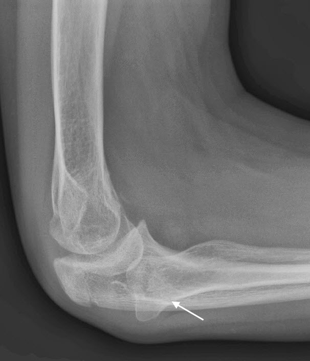
Congenital radial head dislocation is an unusual congenital dislocation in which there is dislocation of the radial head from its usual position. The direction of the displacement of the radial head may be anterior or posterior or lateral.
Congenital radial head dislocation is a rare congenital abnormality of the elbow though statistically most common one.
The dislocation can occur in isolation. But more commonly, it is associated with other conditions or syndromes.
It is bilateral in most of cases.
Congenital dislocation of radial head was described by McFarland.
Pathophysiology
It is often accompanied by radial shortening and bowing. The radial head is hypoplastic [underdeveloped]. The condition can occur in isolation but is often associated with syndromes such as nail-patella syndrome.
In some cases, congenital dislocation of the radial head is genetically transmitted.
Following characteristics are used to differentiate congenital dislocation of radial head from traumatic dislocation
- No history of injury
- Bilateral involvement
- Convex radial head
- Underdeveloped capitellum
- Often present with other congenital anomalies
Elbow is an important joint for crucial for activities of daily living. It acts as a lever arm when positioning the hand functions as a fulcrum for forearm The elbow joint includes the following
- Ulnohumeral joint – Joint between ulna and humerus
- Radiocapitellar joint- Joint between radial head and capitellem
- Proximal radioulnar joint – Joint between the radial head and upper part of ulna
Congenital dislocation of radial head leads to loss of articulation of radicapitellar and proximal radioulnar joints.
Congenital radial head dislocation is posterior in about 70 percent cases is the most common type. Anterior and lateral dislocations are less common making about 15% each.
Causes and Associated Conditions
The exact mechanism of congenital dislocation of radial head is not clear. It is hypothesized that there is a failure of development of a normal capitellum. this failure leads to causing loss of contact pressure required for normal development of the radial head resulting in the malformation of the radiocapitellar joint. There is altered altered biomechanics at the proximal radioulnar joint and abnormal development of the ulna as well.
Congenital dislocation of radial head is known for its associations with many conditions though it can occur in isolation as well. Following conditions are commonly seen to be associated –
- Apert syndrome
- Klinefelter syndrome
- Ehlers-Danlos syndrome
- Nail-patella syndrome
- Klippel-Feil syndrome
- Scoliosis
- Anomalies of lower limb
Clinical Presentation
Congenital dislocation of the radial head is often not noted until the child is 4 or 5 years old. Some cases are not diagnosed till adolescence.
Most of the cases are asymptomatic in the beginning.
Painless limitation of motion and deformity are the main complaints.
The condition may be detected when the patient is evaluated for some other ailment or minor injury.
On examination, the radial head is prominently felt out of its socket. In normal elbow examination, radial head is palpated just below the lateral epicondyle of the humerus and confirmed by supination and pronation of the arm as it rotates during those movements.
Most of the dislocations are felt posteriorly, about 1/ 3 are anterior or lateral.
The ulna is bowed, the direction of convexity depending on the type of dislocation
- Anterior dislocation-ulnar bow is forward
- Posterior dislocation-backward
- Lateral dislocations the ulna will bend laterally.
In anterior dislocations the range of elbow flexion is limited and the radial head may be palpated in the cubital fossa. In posterior dislocations, the elbow will not extend fully and the prominent radial head may be palpated posteriorly.
The range of motion is affected especially that of supination and pronation.
Imaging
X-rays are the basic investigations and often enough for diagnosis. Anteroposterior and lateral views of elbow are done for evaluation of the elbow in congenital dislocation of radial head.
The dislocation might be quite obvious especially in the lateral view. The following tests may help to diagnose the dislocation in cases where there is doubt.
In a normal elbow, a line drawn through the longitudinal axis of the radial shaft bisects the capitellum of the humerus. This normal finding is absent in this condition and suggests a dislocation.
The head of the radius is dome-shaped on its superior surface.

The radius may be short and bowed.
It is important to distinguish congenital dislocation of radial head from traumatic ones.
Following injury patterns are known to cause traumatic dislocation of radial head-
The types of injury that cause traumatic dislocation of the radial head are missed
- Monteggia fracture-dislocations
- Fracture of the radial neck
- Pulled elbow
- Traumatic dislocation of the radial head
Mcfarland proposed the following diagnostic criteria for congenital dislocation of the radial head.
- Absent or hypoplastic and flattened capitellum
- Prominent ulna epicondyle
- Partially abnormal trochlea
- Domed radial head articular surface with a long neck
- Negative ulnar variance
Treatment of Congenital Dislocation of Radial Head
Nonoperative treatment is the first line of treatment of congenital dislocation of radial head. Symptoms are managed as per individual presentation though most of the patients do not need any form of treatment till adolescence when the symptoms get more pronounced.
Surgery is considered in adulthood or till the child attains skeletal maturity.
The following are considered indications for surgical intervention
- Pain in elbow
- Restricted motion of the elbow
- Cosmetic concerns
Radial head excision is the well-established procedure for the treatment of this condition. It can improve appearance but often does not affect the motion
The surgery should be done after the child has attained skeletal maturity as the resection of the radial head in the child results in regrowth of the head and marked proximal migration of radius can occur.
Image Credit: Congenitalhand.wustl.edu