• Skip to main content
  • Skip to primary sidebar
  • Skip to footer
  • General Ortho
  • Procedures
  • Spine
  • Upper Limb
  • Lower Limb
  • Pain
  • Trauma
  • Tumors
  • Newsletter/Updates
  • About Us
  • Contact Us

Bone and Spine

Orthopedic health, conditions and treatment

Coxa Vara Causes and Treatment

By Dr Arun Pal Singh

In this article
    • Relevant Anatomy
    • Classification of Coxa Vara
      • Associations
    • Clinical Presentation
    • Imaging
      • X-rays
      • CT
    • Treatment of Coxa Vara
    • Congenital Coxa Vara

Coxa vara describes a deformity of the hip where the angle formed between the head and neck of the femur and its shaft is decreased, usually defined as less than 120 degrees. This angle is also known as Mikulicz angle.

It can be congenital or acquired. The common mechanism in congenital cases is a failure of medial growth of the physeal plate.

As coxa vara is a symptom of many diseases, it is usually classified according to the condition which causes it.

Relevant Anatomy

Read about proximal femoral anatomy here.

Adult femoral shaft and femoral neck make an angle that ranges between 120 degrees to 140 degrees.

If this angle is more than 140 degrees, one is said to have coxa valga. Conversely, a decrease in this normal neck—haft angle to <120 degrees, coxa vara is said to be present.

Coxa vara and coxa vlaga
Image: Wikipedia

In anatomical positions, the neck is depressed than normal. The depression of the neck results in a certain mechanical disadvantage. The normal apposition between the joint surfaces is lost since the head of the femur no longer accurately fits the acetabulum.

The trochanter is displaced upwards, and hence, during the abduction, is liable to imping on the side of the pelvis.

The marked shortening of the limb leads to a waddling gait, not unlike that of congenital dislocation of the hip.

Classification of Coxa Vara

  • Idiopathic:
    • Congenital – mild or severe coxa vara, with associated congenital anomalies
  • Developmental –  progressive, usually appearing between 2-6 years
    • Mucopolysaccharidoses (e.g. Morquio’s disease)
    • Dysplasia epiphysealis multiplex
    • Cleidocranial dysostosis
  • Rachitic –  usually associated with active rickets
  • Adolescent: secondary to slipped capital femoral epiphysis
  • Traumatic – usually following fracture of the femoral neck (rare in children)
  • Inflammatory –  secondary to tuberculosis or other infection
  • Secondary to other underlying bone diseases such as:
    • Osteogenesis imperfecta
    • Cretinism
    • Dyschondroplasia
    • Paget’s disease
    • Osteoporosis
    • Fibrous dysplasia
    • Severe osteoarthritis
    • Legg-Perthes’ disease

Associations

  • Congenital dislocation of hip – developmental hip dysplasia
  • Faulty development of the femur
  • Craniocleidodysostosis
  • Camptodactyly arthropathy coxa vara pericarditis  syndrome

Clinical Presentation

The patient is brought because of abnormal painless gait or limp. Patient walks with  Trendelenburg limp in unilateral coxa vara and a waddling gait in bilateral coxa vara.

Following findings can be seen on examination

  • Limb length discrepancy
  • Prominent greater trochanter
  • Limitation of abduction and internal rotation of the hip.
  • Patients may also show femoral retroversion or decreased anteversion.

Imaging

X-rays

hilgenreiner-epiphyseal-angle
Hilgenreiner Epiphyseal-Angle, Image: Clinical Gate

The recommended views are anteroposterior view of the hip with limb internally rotated.

Findings are

  • Neck-shaft angle <120 degrees
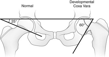
  • Increased Hilgenreiner’s epiphyseal angle (normal <25 degrees)
    • Determined on AP view of the pelvis as the angle between Hilgenreiner’s line [Line drawn horizontally through the inferior aspect of both triradiate cartilages.] and a line through the proximal femoral physis
  • Triangular metaphyseal fragment in the inferior femoral neck. It appears as inverted-Y radiolucency
  • Decreased femoral anteversion
bilateral coxa vara
Bilateral coxa vara

CT

  • CT along with 3D image reconstruction is done to plan for surgery, mark proximal femur defects and gauge the orientation of deformity.
  • It helps to find deformity configuration, bone stock, and physeal widening

Treatment of Coxa Vara

Treatment objective is to restore the neck-shaft angle so that the epiphysial plate is restored to decrease shear forces and promote ossification of the femoral neck defect. The treatment is corrective osteotomy.

Indications for surgical intervention are

  • Neck – shaft angle < 90 degrees
  • Progressive  deformity.
  • Vertical physis and a significant limb length discrepancy
  • HE angle> 60 degrees

Patients with HE angle 45 – 60 degrees is closely followed up. Patients with HE angle < 45 degrees are known to have spontaneous resolution.

A valgus osteotomy reduces the deformity, normalize the neck-shaft angle, improves the gluteal forces, improves joint congruency and increases limb length to reduce the limb length discrepancy.

Congenital Coxa Vara

Coxa vara may be primary congenital deformity or may occur in association with other congenital.

The patient is usually small in stature and limps, and, in bilateral cases, rather resembles a patient with congenital dislocation. Often there is pain and stiffness. On examination, the greater trochanter is on a level higher than the normal, with consequent shortening, while there may be a flexion contracture present.

Radiographic features are

  • The angle of the neck is reduced to something below a right angle.
  • The neck varies in length but is short and may even be non- existent
  • The head is unusually translucent and may be fluffy in outline.
  • Acetabular deformity due to the malposition of the head.
  • In extreme cases, greater trochanter curves inwards and may articulate with the ilium above the acetabulum.

Untreated, it can result in severe deformities and disability.

Treatment is a corrective osteotomy. This involves a wedge removal with the base on the lateral side.

Spread the Knowledge
  • 2
    Shares
  •  
    2
    Shares
  • 2
  •  
  •  
  •  
  •  

Filed Under: Lower Limb

About Dr Arun Pal Singh

Arun Pal Singh is an orthopedic and trauma surgeon, founder and chief editor of this website. He works in Kanwar Bone and Spine Clinic, Dasuya, Hoshiarpur, Punjab.

This website is an effort to educate and support people and medical personnel on orthopedic issues and musculoskeletal health.

You can follow him on Facebook, Linkedin and Twitter

Reader Interactions

Comments

  1. M. Khalid says

    April 24, 2019 at 4:56 pm

    Hi doctor sb

    Does any plaster require after surgery. Please also brief about estimated time require to heal up the surgery wounds and after how much time 2nd side can be operated

  2. Dr Arun Pal Singh says

    May 1, 2019 at 9:23 pm

    Khalid,
    The use of plaster would depend on the fixation and its strength. For other questions, your treating doctor would be in better position to answer.

Leave a Reply Cancel reply

Your email address will not be published. Required fields are marked *

This site uses Akismet to reduce spam. Learn how your comment data is processed.

Primary Sidebar

Enchondromatosis or ollier disease upper limb

Ollier Disease – Causes, Presentation and Treatment

Ollier’s disease is a rare, nonhereditary skeletal disorder characterized by a multifocal intramedullary proliferation of hypercellular dysplastic cartilage. It is a type of enchondromatosis. Maffucci’s syndrome is another type of enchondromatosis where the skeletal lesions are accompanied by hemangiomas of skin, soft tissu, and viscera. Enchondromatosis represents a distinct developmental disorder of enchondral ossification and […]

Internal fixation of Distal TIbia Plate In Fractures of Distal Tibia

Internal Fixation of Fractures – Principles and Devices

Internal fixation of the bone is a type of fixation where the fixing device is not exposed to outside environment as it is buried under the soft tissue. This is in contrast to external fixation. Bone acts as a supporting structure and transmits load. Different forces that act on bone are compression, tension and shear. […]

Incomplete spinal injuries

Incomplete Spinal Injury Patterns

An incomplete spinal injury is defined as spinal cord injury with some preserved motor or sensory function below the injury level. The spared function may be  including Voluntary anal contraction (sacral sparing) Palpable or visible muscle contraction below the injury level Perianal sensation present It must be noted that the completeness of the lesion is the […]

Lenke classification system for scoliosis

Lenke Classification System for Scoliosis

Lenke classification is a comprehensive radiographic classification of Adolescent Idiopathic Scoliosis named after Dr Lawrence Lenke who was instrumental in devising it. Adolescent idiopathic scoliosis is the most common types of scoliosis encountered. Though originally intended to be include factors in 3 planes, it eventually included two saggital and coronal plane. The classification was published […]

Normal supination of Foot Movement

Supination of Foot and Oversupination

Supination of the foot is the natural movement of the foot as it rolls out during the gait cycle. Supination of foot is the movement of the subtalar joint (between the talus and calcaneus)  and consists of three movements – inversion, plantar flexion, and adduction. Inversion – Sole of the foot is turned inwards Plantar […]

Calluses in hand

Interesting Names for Common Calluses

Callus is hyperkeratotic thickening due to repetitive friction. Presentation of calluses and corns are discussed here. In this article, I am writing about the commonly used terms for different types of calluses. These are vernacular terms and are interesting to note as they try to convey the etiology. Jeweler’s callus or  Cherry Pitter’s thumb or […]

Ulnar gutter splint

Ulnar Gutter Splint Application Procedure

An ulnar gutter splint extends from the proximal forearm to distal interphalangeal joints of fingers on the ulnar aspect of fingers wrist and forearm and is applied for various injuries of the ulnar aspect of the wrist and hand. It is also called Boxer splint. The splint is applied on the ulnar aspect and conforms to […]

Browse Articles

Footer

Pages

  • About
    • Policies
    • Contact Us

Featured Article

Digital Block Procedure – Indications and Techniques

Digital nerve blocks are used for numbing digits [fingers, toes] for a procedure on the digits. Injury and infection are the two most common reasons … [Read More...] about Digital Block Procedure – Indications and Techniques

Search Articles

© Copyright: BoneAndSpine.com