Last Updated on September 25, 2023
Diffuse idiopathic skeletal hyperostosis also called Forestier’s disease is characterized by unique, flowing (wave-like in shape) calcification along the sides of the vertebrae of the spine. It is also commonly associated with inflammation and calcification of tendons at their attachment points to bone, a condition called enthesitis. Enthesitis can frequently lead to bone spurs such as heel spurs.
DISH usually presents with back pain and spinal stiffness.
It occurs in 6-12 percent of people and is not common before 50 years of age. Females are affected slightly more than males.
Though it can occur anywhere in the spine, it is more common in the thoracic spine, followed by the lumbar and cervical spine.
The facet joint and sacroiliac joints are not involved in diffuse idiopathic skeletal hyperostosis.
Cause of Diffuse Idiopathic Skeletal Hyperostosis
At present no cause is known. There is a tendency toward ossification of ligament, tendon, and joint capsule. These are also broadly termed as enthesial insertions.
The various known risk factors are
- Obesity
- Hypertension
- Metabolic syndrome
- Type 2 diabetes mellitus
- Hyperuricemia
Clinical Presentation
Diffuse idiopathic skeletal hyperostosis is a completely asymptomatic phenomenon.
The condition is discovered inadvertently.
However, there are reported presentations and most of these symptoms arise due to impingement by bony overgrowth.
In the back, the patient may complain of mild chronic back pain and stiffness. The symptoms could be worse in the morning or in cold weather, that could be worse in the morning.
Other symptoms can be
- Pain in the joints
- Neck pain
- Difficulty in swallowing- dysphagia (Due to neuropathy or impingement)
There is an increased susceptibility to spinal fractures because of the ankylosis of the spine.
Diagnosis
No laboratory tests are indicated.
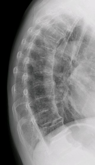
X-rays
AP and lateral views of the spine are the recommended views. The usual findings suggestive of DISH are syndesmophytes at 3 successive levels (4 contiguous vertebrae).
- Thoracic spine
- Involved almost in all cases
- Right-sided involvement
- Absence on one side of the thoracic spine is thought to be due to the influence of aortic pulsations.
- T7-T11 are particularly involved
- Cervical spine
- Bone formation on the anterior side
- Disc spaces are preserved (versus ankylosing spondylitis where disc spaces would be ossified)
- Lumbar spine
- Symmetrical syndesmophytes (compare with one-sided involvement of thoracic)
- Peripheral joints – will show degenerative changes
- Other changes
- Ossification of the nuchal ligaments in the skull
- Enthesopathy at the ischial tuberosities
- ossification of the sacrotuberous ligament and symphysis pubis
- Ossification of the quadriceps and infrapatellar tendons
- Ossification of the Achilles tendon and the plantar aponeurosis
- Ossification of the triceps tendon
- Subcutaneous calcification
Radiographs demonstrate typical changes in the spine as discussed earlier in this article. Other findings in other regions may be
Recognition of diffuse idiopathic skeletal hyperostosis facilitated by its separation from the body of the vertebrae. This gives rise radiologically to the appearance of a radiodense line paralleling the longitudinal axis of the spine but separated by a clearly definable space.
Ossification at sites of the tendon, ligamentous, or joint capsule insertion (enthesitis) is strongly suggestive of the diagnosis. Enthesial reaction at the iliac crest and ischial tuberosities, quadriceps insertion, metacarpal, and phalangeal capsule insertion may be present.
Usually, the X-rays are sufficient to reach the diagnosis.
CT or MRI are indicated in patients with a history of trauma to evaluate for the presence of occult fractures.
Computed tomography may also be performed to evaluate complications, such as fracture, or to conform pressure effects on the trachea, esophagus, and veins.
Criteria for Diffuse Idiopathic Skeletal Hyperostosis
- Resnick and Niwayama
- Flowing calcifications and ossifications along the anterolateral aspect of at least 4 contiguous vertebral bodies, with or without osteophytes
- Preservation of disc height in the involved areas and an absence of excessive disc disease
- Absence of bony ankylosis of facet joints and absence of sacroiliac erosion, sclerosis, or bony fusion.
- Julkunen et al
- Definite
- Bridging of four contiguous vertebral bodies
- Minimal intervertebral disc disease
- No facet joint ankylosis
- Probable
- Bridging of two contiguous vertebral bodies
- Bilateral patellar tufting
- Heel spur
- Olecranon tufting.
- Possible [Either of two situations]
- Two vertebrae joined and no extraspinal enthesophytes
- Symmetrical extraspinal enthesophytes without any spinal involvement
- Definite
Unlike ankylosing spondylitis, Diffuse idiopathic skeletal hyperostosis does not involve the sacroiliac joint.
The most commonly involved spine is lower thoracic spine involvement which is also typical of diffuse idiopathic skeletal hyperostosis, but the lumbar and cervical spine also can be affected. The right side of the spine is more commonly involved. The lesser involvement of the left side of the spine is attributed to the pulsation of the aorta on this side.
Treatment
There is no specific treatment and the patients are treated symptomatically with anti-inflammatory drugs, local heat, and physical therapy. Any complication is treated accordingly.
Nonoperative treatment
- Modification of activities of daily life
- Physical therapy
- Use of orthotics like brace
- Drugs to relieve pain
Operative Treatment
Surgery is done to relieve the spinal cord of any compression. Stabilization often accompanies spinal decompression.
Some conditions that may require decompression are
- Lumbar stenosis
- Cervical myelopathy
- Deformity that warrants correction
Complications of Diffuse Idiopathic Skeletal Hyperostosis
- Posterior longitudinal ligament ossifications may impinge on the spinal cord on rare occasions.
- Reduced vertebral column flexibility predisposes to vertebral fracture.
- Diffuse idiopathic skeletal hyperostosis is not a source of morbidity or mortality. However, increased mortality is noted in cervical injury in DISH
References
- Nascimento FA, Gatto LA, Lages RO, Neto HM, Demartini Z, Koppe GL. Diffuse idiopathic skeletal hyperostosis: A review. Surg Neurol Int. 2014. 5 (Suppl 3):S122-5. [Link].
- Luo TD, Varacallo M. Diffuse Idiopathic Skeletal Hyperostosis (DISH). 2020 Jan. [Link]
- Kaffel D, Kchir H. Dysphagia related to diffuse idiopathic skeletal hyperostosis (DISHphagia). Clin Case Rep. 2019 Nov. 7 (11):2265-2266 [Link]