• Skip to main content
  • Skip to primary sidebar
  • Skip to footer
  • General Ortho
  • Procedures
  • Spine
  • Upper Limb
  • Lower Limb
  • Pain
  • Trauma
  • Tumors
  • Newsletter/Updates
  • About Us
  • Contact Us

Bone and Spine

Orthopedic health, conditions and treatment

Digital Xray Radiogrammetry for Bone Mineral Density

By Dr Arun Pal Singh

In this article
    • Digital Xray Radiogrammetry

Radiogrammetry is a technique that uses a conventional radiograph for the assessment of bone status by using cortical width as a measure of bone strength.

Osteoporosis is defined as a systemic skeletal disease characterized by low bone mass and micro-architectural deterioration of bone tissue, with a consequent increase in bone fragility and susceptibility to fractures.

Measurement of bone mineral density forms the basis for the working definition of osteoporosis.

Fractures are the hallmark of osteoporosis. The risk of fracture in osteoporosis is determined by the risk of fall, the force of impact and the strength of bone. Reduced bone mineral density is an important component of fracture risk in osteoporosis.

digital-xray-radiogrammetry

Following methods are used for measuring  bone mineral density

  • Radiography
  • Radiogrammetry
  • Radiographic absorptiometry
  • Single- (SXA) and dual X-ray absorptiometry (DXA)
  • Spinal and peripheral quantitative computed tomography (QCT/ pQCT)
  • Quantitative ultrasound (QUS)
  • Magnetic resonance imaging.

In this article, we would discuss radiogrammetry and digital Xray radiogrammetry.

Digital Xray Radiogrammetry

Digital x-ray radiogrammetry uses computerized radiogrammetric analysis and textural analysis of a digitized radiograph of the hand. Information from the middle three metacarpals is used by the system to generate a BMD estimate. Digital Xray Radiogrammetry was approved by US FDA in 1999.

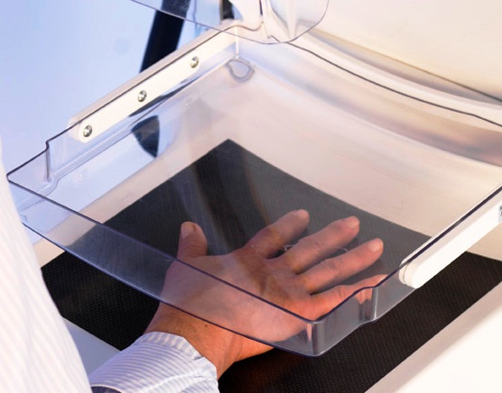
A plain radiograph of the non-dominant hand (preferably) is taken under standardized conditions. After checking the quality of the radiograph, it is placed in a scanner, which digitizes the image. The initial computer processing identifies anatomical landmarks and automatically defines the ‘Regions of Interest’ (ROIs). The digitized image is displayed on the screen with the ROIs superimposed. There is no operator activity involved in placing the regions.

The system computes the Digital x-ray radiogrammetry BMD [DXR-BMD] based on the cortical thickness of the ROIs. About one centimeter of both the cortices in ROI is studied. The cortical thickness is measured 118 times per vertical centimeter of the ROI in each cortex. Thus a total of 236 measurements per centimeter are made over the two cortices.

The resulting  DXR-BMD estimate is presented as

  1. Absolute bone mineral density estimate in g/cm 2
  2. Graphic form, in a DXR-BMD versus age plot, together with a reference curve
  3.  T score
  4. Z score.

Repeated studies have shown a  high correlation of DXR -BMD with DXA-BMD. It is said to be good in predicting fractures as well as discriminate patients with and without fractures.

Moreover, DXR-BMD can also detect a change in bone density with time and anti-resorptive therapy.

The investigation is operator independent.

Spread the Knowledge
  • 7
    Shares
  •  
    7
    Shares
  • 1
  • 6
  •  
  •  
  •  

Filed Under: Imaging

About Dr Arun Pal Singh

Arun Pal Singh is an orthopedic and trauma surgeon, founder and chief editor of this website. He works in Kanwar Bone and Spine Clinic, Dasuya, Hoshiarpur, Punjab.

This website is an effort to educate and support people and medical personnel on orthopedic issues and musculoskeletal health.

You can follow him on Facebook, Linkedin and Twitter

Reader Interactions

Leave a Reply Cancel reply

Your email address will not be published. Required fields are marked *

This site uses Akismet to reduce spam. Learn how your comment data is processed.

Primary Sidebar

Dorsal view of polydactyly

Polydactyly or Supernumerary Digit – Presentation and Treatment

Polydactyly or supernumerary digit refers to the presence of means extra digits in the hand or foot. It is also called hyperdactyly. It is the most common congenital digital anomaly of the hand and foot. It may appear in isolation or in association with other birth defects and syndromes. The sporadic polydactyly is often autosomal […]

prepatellar bursitis image

Prepatellar Bursitis or Housemaid Knee

Prepatellar bursitis is inflammation and swelling of the prepatellar bursa which lies between the skin and the patella separating the patella from the patellar tendon and skin. Normally, it contains a minimal amount of fluid and is not appreciable but with inflammation, it could swell substantially. Prepatellar bursitis is also known as the beat knee, […]

Fracture of scaphoid

Wrist Biomechanics and Mechanism of Wrist Injuries

Wrist biomechanics and function of the human wrist joint are unique. The wrist joint is a diarthrodial joint composed of two rows of carpal bones. The proximal row articulates with the distal articular surface of the radius, which consists of the scaphoid fossa and the lunate, fossa. The distal surface of the scaphoid, lunate, and […]

Anterior Spinal Artery Syndrome

Anterior Spinal Artery Syndrome

Anterior spinal artery syndrome affects the supply of the anterior spinal artery which supplies the ventral two-thirds of the spinal cord and Medulla. Infarction or blockage of this artery jeopardizes the supply to the cord and results in symptoms of weakness and loss of pain and temperature sensation below the level of injury. Causes of […]

Sprengel Deformity

Sprengel Deformity – Congenital High Scapula

Sprengel deformity is a congenital condition where the scapula is small and located higher than its normal position because of its failure to descend to its position. There could also be scapular winging and presence of omovertebral bone 30-50%]. Omovertebral bone is an anomalous bone connecting the elevated scapula to the cervical spine. It is […]

Grades of Symphalism

Symphalangism – Causes, Classification and Treatment

Symphalangism is a condition where there is congenital ankylosis [fusion] of the interphalangeal joints  of the phalanges of the hand or foot. The condition may involve metacarpophalangeal joints as well. The condition occurs because of the failure of the interphalangeal joint to differentiate during the development. Symphalangism leads to the functional impairment and unsightliness of […]

Illustration of Posterior cervical spine surgery

Cervical Spine Surgery in Disc Disease

Cervical spine surgery for is most commonly performed for radiculopathy and cervical degenerative disc disease. The goal of cervical spine surgery is similar to lumbar spine surgery – adequate decompression of the nerve roots and stabilization of the spine Anterior cervical discectomy Anterior cervical discectomy  and fusion Anterior cervical discectomy  and fusion with internal fixation […]

Browse Articles

Footer

Pages

  • About
    • Policies
    • Contact Us

Featured Article

Flail Chest Injury – Causes, Presentation and Treatment

Flail chest is caused by severe blunt injury which causes multiple [3 or more] segmental [rib fractures at two points say anterior and posterior] rib … [Read More...] about Flail Chest Injury – Causes, Presentation and Treatment

Search Articles

© Copyright: BoneAndSpine.com