Last Updated on November 20, 2023
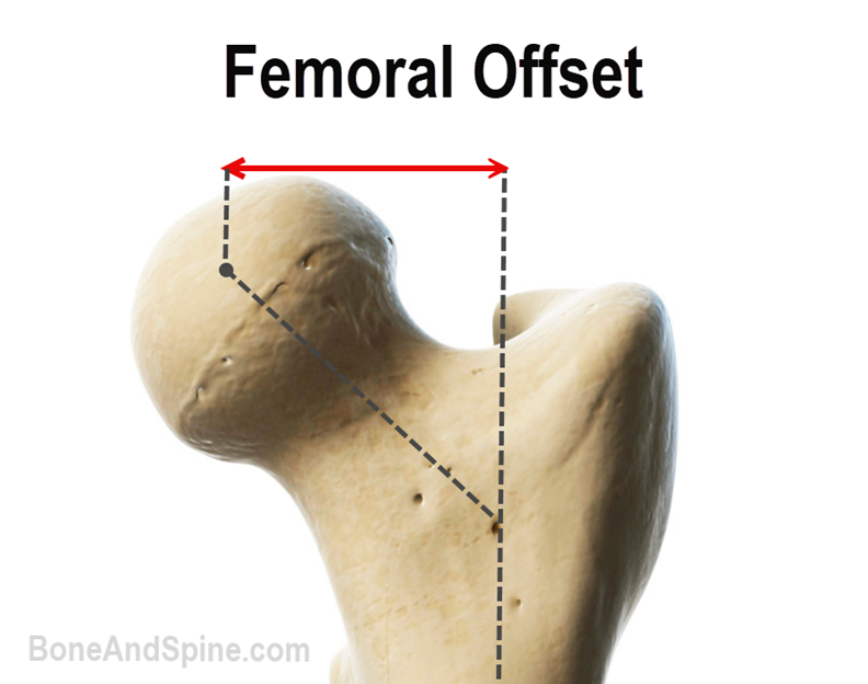
Femoral offset is the distance from the center of rotation of the femoral head to a line dissecting the long axis of the femur. In the case of total replacement of the hip, the offset is considered as the distance from the center of rotation of the femoral head to a line bisecting the long axis of the stem.
Normal femoral offset varies between 30 and 60 mm.
Acetabular Offset and Global Offset
Conventionally, femoral offset has been measured on radiographs. A computed tomography scan is more accurate than plain radiography. Acetabular offset and global offset are other important parameters along with femoral offset.
- Fem Offset: The distance from the center of rotation of the femoral head to a line dissecting the long axis of the femur.
- Acetabular Offset: It is the distance between the center of the femoral head and the true floor of the acetabulum.
- Global Offset: The sum of both – femoral and acetabular offsets- is called global offsets.
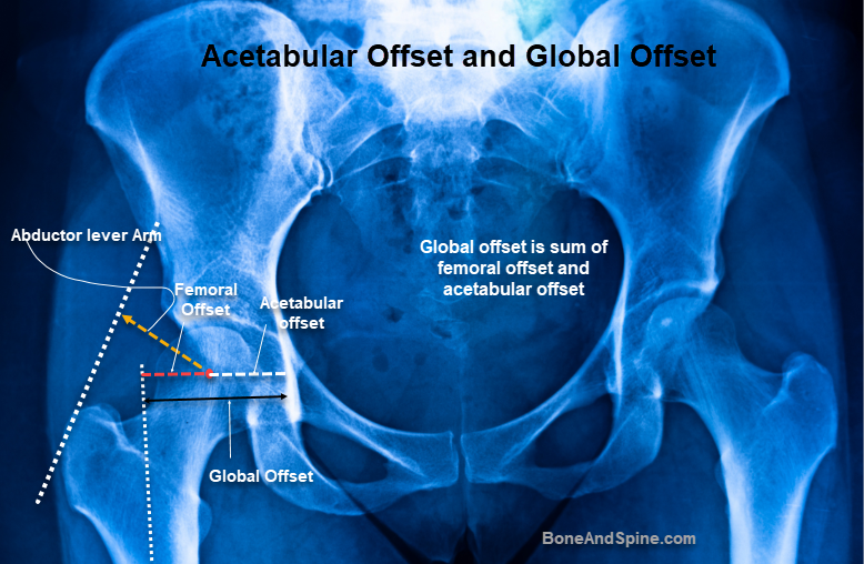
- Abductor Muscle Arm: The path of the abductor muscles might be represented by drawing a line tangential to the lateral margin of the greater trochanter. The abductor muscle lever arm is thus perpendicular to it
Significance of Femoral Offset
Studies have shown a strong correlation between
- Femoral offset
- Abductor lever arm
- Hip abductor strength
Therefore, restoration of the offset is essential. This improves the function as well as provides a longer life to the hip arthroplasty.
The concept of offset is very important for total hip arthroplasty prosthesis design and choice of implant for a particular individual.
- A decrease in femoral offset would move the femur closer to the pelvis medially.
- This can lead to impingement of greater trochanter in extremes. The medial movement would also result in soft tissue relaxation. Both of these factors can lead to instability of the implant and possible dislocation.
- Moreover, when the offset decreases, greater force is required by the abductor’s muscles to balance the pelvis, and the resultant force across the hip joint also increases resulting in greater wear and tear.
- An increase in femoral offset moves the femur laterally resulting in decreased chances of impingement, better tension in soft tissues, and better stability.
- It decreases the force required by the abductor muscles to balance the pelvis, which will improve gait. As well, the resultant force decreases with increased offset, which may result in less wear and loosening over time.
A change in femoral offset does not affect the leg length and thus provides a measure to make an adjustment without altering the leg length.
In total hip replacement surgery, the offset needs to be determined preoperatively for better planning of the surgery.
The femoral neck-shaft angle is responsible for the anatomical fem offset whereas femoral neck anteversion defines the physiological offset. An increase in anteversion leads to a decrease in the functional offset.
The medialization of the hip center of rotation provides a better abductor lever arm and better acetabular coverage. A prosthesis with lateralized femoral stems allows increased offset that is required for abductor lever arm restoration in these cases. Every individual is different and each case of arthroplasty should be evaluated and planned on basis of anatomical variations and physiological requirments.
References
- Yamaguchi, M. Naito, I. Asayama, T. Ishiko. Total hip arthroplasty: the relationship between posterolateral reconstruction, abductor muscle strength, and femoral offset. J Orthop Surg, 12 (2004), pp. 164-167
- T.J. Spalding, Effect of femoral offset on motion and abductor muscle strength after total hip arthroplasty. J Bone Joint Surg (Br), 78 (1996), p. 997
- Sakalkale DP, Sharkey PF, Eng K, Hozack WJ, Rothman RH. Effect of femoral component offset on polyethylene wear in total hip arthroplasty. Clin Orthop Relat Res. 2001;388:125–134. [link]