Last Updated on August 2, 2019
Fibular hemimelia or congenital absence of fibula is now known as a fibular deficiency. It causes anteromedial bowing of tibia.
But it is still popularly known as fibular hemimelia and is characterized by the absence of fibula varying from minimal shortening of the fibula to its complete absence.
Fibular hemimelia is rare and expression may range from mild deformity to severe deformity.
The exact cause is unknown though interference with limb-bud development has been suggested to plays an important role.
To support this theory, it is held that other associated abnormalities of the knee, leg, ankle, and foot also are related to the fibular field and may occur in association.
Other terms for fibular hemimelia, postaxial hypoplasia of the lower extremity, encompasses all these abnormalities too
Clinical Presentation of Fibular Hemimelia
Clinical presentation of fibular hemimelia ranges from an absent fifth toe in a newborn or a difference in limb lengths to severe fibular deformities.
Dimpling of skin over midanterior tibia may be seen. Foot may be in equinovalgus position.
Associated abnormalities may be present which could be at birth or evolve with growth. These are
- Proximal femoral focal deficiency
- Coxa vara
- Femoral hypoplasia with external rotation
- Lateral subluxation of patella
- Hypoplastic lateral femoral condyle [may cause genu valgum]
- Flattened tibial eminence with absent cruciate ligament
- Short or bowed tibia
- Ball and socket ankle
- Absent tarsal bones
- Tarsal coalitions
- Absent foot rays
It is essential to evaluate the entire limb so that everything could be factored in for developing a treatment plan.
Imaging
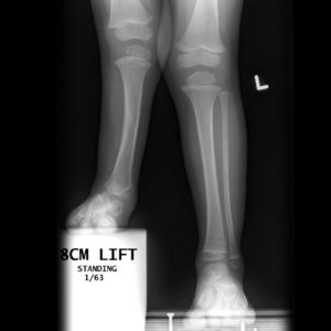
Image Credit: Radiopaedia
X-rays of the whole limb in standing position are preferable to assess the deformity and associated conditions. Opposite extremity is imaged for control and comparison.
Whenever required additional x-rays can be done for detailed analysis.
Classification of Fibular Hemimelia
Classification is based on x-rays
The Achterman and Kalamchi classification:
Type IA
The proximal fibular epiphysis is distal to the level of the tibial growth plate with the distal fibular epiphysis proximal to the talar dome
Type IB
The proximal fibula is absent for 30-50% of its length; the distal fibula is present but does not adequately support the ankle (see the second and third images below)
Type II
The fibula is completely absent
MRI
It better-assessed bone and soft tissue abnormalities of the knee joint in patients with fibular hemimelia
Treatment of Fibular Hemimelia
Treatment is mostly surgical. Minor deformities can be left as such and observed.
The goal of the surgical treatment is to provide a maximal function by maintaining alignment, length, and stability of the involved lower limb. This goal may not always be achievable though and if it is so amputation followed by a functional prosthesis should be considered. For example, a patient with non-functional foot should undergo amputation.
The limb-length discrepancy is one of the more difficult to address. If limb length discrepancy
- <10% – lengthening of the involved limb or contralateral epiphysiodesis
- > 30% – amputation
- 10-30% (the most challenging group) – Lengthening of the involved limb with a contralateral epiphysiodesis to inhibit the growth
If amputation is determined to be appropriate, the unmodified Syme amputation is generally recommended.
References
- Stevens PM, Belle RM. Screw epiphysiodesis for ankle valgus. J Pediatr Orthop. 1997 Jan-Feb. 17(1):9-12.
- Shabtai L, Specht SC, Standard SC, Herzenberg JE. Internal lengthening device for congenital femoral deficiency and fibular hemimelia. Clin Orthop Relat Res. 2014 Dec. 472(12):3860-8.
- Das S, Ganesh GS, Pradhan S, Mohanty RN. Outcome of eight-plate hemiepiphysiodesis on genu valgum and height correction in bilateral fibular hemimelia. J Pediatr Orthop B. 2014 Jan. 23(1):67-72.
- Pappas AM, Hanawalt BJ, Anderson M. Congenital defects of the fibula. Orthop Clin North Am. 1972 Mar. 3(1):187-99.
- Manner HM, Radler C, Ganger R, Grill F. Knee deformity in congenital longitudinal deficiencies of the lower extremity. Clin Orthop Relat Res. 2006 Jul. 448:185-92.