Last Updated on August 6, 2023
What are Flexion-Extension Xrays of Cervical Spine
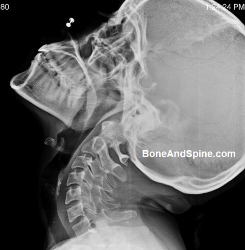
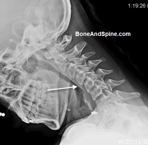
Flexion-extension xrays of cervical spine are dynamic x-ray studies of the neck. The cervical spine flexion-extension x-rays are taken in the lateral position and show seven cervical vertebrae and their relations.
These x-rays of the cervical spine are helpful in finding instability of the cervical spine that is not very obvious in routine images. It can also be used to assess the healing response of cervical spine injuries and other pathologies.
Flexion-extension xrays of cervical spine are taken in different positions of the neck i.e. flexion and extension. This is called dynamic radiography. The other kind that is routinely done by taking different views of the cervical spine is called static radiography.
Sometimes, in dynamic radiography, additional distraction forces may be applied by putting traction and the displacement may be studied. Any abnormal displacement indicates insufficiency.

The x-ray above and below are of 38 years old lady who suffered from chronic neck pain. Her routine x-ray of the cervical spine revealed a kyphotic deformity at C4-C5 level. Flexion-extension x-rays of cervical spine views were done. While the deformity got corrected in extension view, it got exaggerated in the flexion suggesting dynamic instability. [Images above and below]

Indications
When are Flexion-Extension Xrays of Cervical Spine Done?
Cervical Spine Fexion-extension xrays are done to evaluate spinal instability. This is the main indication. It can also be done in acute trauma of the spine where routine films are normal, to assess the ligamentous injury. Bu with better imaging investigations available, these are done less frequently in acute trauma to avoid patient inconvenience and risk of further injury.
Contraindications
When Flexion-Extension Xrays of Cervical Spine are Not Done?
- – When the patient has an altered state of consciousness
- Head injury
- Intoxicated individual
- Patients with neurologic deficit
- Patient is not able to flex or extend the neck
Procedure
- Position – Erect, and the patient’s neck is positioned for a lateral view of the cervical spine. The patient may sit or stand depending on the machine and convenience of the patient
- The x-rays are taken in various degrees of flexion [chin down] and extension [chin up]of the cervical spine as required. A demonstration beforehand would make the procedure easy to manage.
- The exposure focus is around C4 about 2.5 cm above the jugular notch
- A good image would enable visualization of C7 to T1.
- The image is labeled as ‘flexion’ or ‘extension’.
- A good flexion image should demonstrate the well-separated spinous process.
- A good extension image shows the crowding of the spinous process.
Inference
An intersegmental translation of more than 2.5 mm is considered to suggest instability of the spine.
Intersegmental translation is found by comparing flexion and extension views by tracing the posterior line of the vertebra.
Detailed analysis of flexion-extension xrays of cervical spine are done as follows
In flexion View
- Atlanto Dens Interval or ADI
- It is the distance between the odontoid process and the posterior border of the anterior arch of the atlas
- In children, it should be less than 3.5 mm
- Adults less than 3 mm
- The cervical spine in flexion should assume gentle kyphosis and there should be break of the curve indicating greater intersegmental motion
- Symmeteric interspinous and interlaminar distances
- There should not be any widening of the facet joint & intervertebral spaces
- No vertebral body angulation or translation
The instability is indicated by
- Disc widening of 1.7 mm or more
- Translation of 3.5 mm or more
In Extension View
- Cervical spine has mild lordosis
- The spine should be in smooth curve and there should not be any angulation or translation
The flexion view is the most helpful in detecting ligamentous injury and determining the integrity of the supporting structures.
Conclusion
Flexion-extension xrays of cervical spine are done when initial investigations suggest near the normal spine or reveal a deformity that needs to be determined as fixed or correctable.
Flexion-extension xrays of cervical spine are also done when there is enough evidence to suggest instability.
The problem can be as a result of trauma or some other disease. In trauma, however, flexion-extension xrays of cervical spine are usually contraindicated for patients with known acute cervical spine fractures and dislocations. They are deferred until a patient has documented the absence of cognitive impairment, has overcome the acute post-injury state and has no apparent signs of spinal trauma.
In fact, better imaging modalities like CT or MRI obviate the need for flexion-extension X-rays of the cervical spine in cases of patients with trauma.
References
- Insko EK, Gracias VH, Gupta R, Goettler CE, Gaieski DF, Dalinka MK. Utility of flexion and extension radiographs of the cervical spine in the acute evaluation of blunt trauma. J Trauma. 2002; 53: 426
doi: 10.1097/00005373-200209000-00005.[Link] - Sanchez B, Waxman K, Jones T et al. Use of flexion and extension radiographs of the cervical spine to rule out acute instability in patients with negative computed tomography scans. J Orthop Trauma. 2011; 25: 51
doi: 10.1097/01.ta.0000171449.94650.81 [Link]