• Skip to main content
  • Skip to primary sidebar
  • Skip to footer
  • General Ortho
  • Procedures
  • Spine
  • Upper Limb
  • Lower Limb
  • Pain
  • Trauma
  • Tumors
  • Newsletter/Updates
  • About Us
  • Contact Us

Bone and Spine

Orthopedic health, conditions and treatment

Four Corner Arthrodesis [Fusion] of the Wrist

By Dr Arun Pal Singh

In this article
    • Procedure
    • Complications of Four Corner Arthrodesis

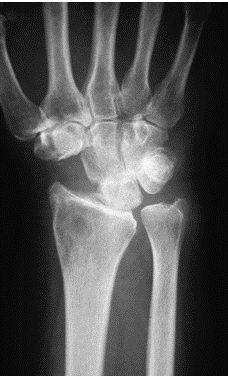
Four corner arthrodesis involves excision of scaphoid and fusion of the remaining wrist bones in neutral alignment. It is motion preserving limited arthrodesis and provides pain relief and improved grip strength.

This four corner arthrodesis is based on the principle that the radiolunate articulation is often spared from degenerative changes from conditions.

Indications

  • SLAC wrist
  • Chronic dynamic carpal instability
  • Chronic perilunar instability not amenable to soft tissue surgery

Contraindications

  • Ulnar Translocation
  • Radiolunate arthritis

Procedure

The surgical procedure is typically performed under regional anesthesia.

A standard operating table and radiolucent hand table is used. The patient is kept in supine position.

A standard dorsal longitudinal incision between the third and fourth extensor compartment of the wrist. The incision is centered on proximal carpal row.

The tubercle of Lister is used as a landmark.

Extensor compartments of forearm
Extensor compartments of Wrist

Divide the third and fourth compartments by incising the retinaculum and then septae. The tendons are retracted unlarwards

The joint capsule is opened individual carpal joints are exposed.  Expose the midcarpal joint and the radial half of the radiocarpal joint.

Split the dorsal dorsal radiocarpal and dorsal intercarpal ligaments taking along the apex of the triquetrum

The flap is elevated radially detaching the dorsal capsule from the radius.

The ulnocarpal joint is exposed by splitting the dorsal radiocarpal ligament longitudinally followed by incising the capsule along the extensor carpi tendon.

This flap is elevated proximally

The scaphoid is excised after identification either piecemeal fashion with a rongeur or sharply using a scalpel. Volar radioscaphocapitate ligament should be carefully protected.

After excision, the wrist is prepared for fusion.

The fusion surfaces between the lunate, capitate, hamate, and triquetrum are then denuded down to the cancellous bone.

Volar third cartilage is removed from lunate and capitate to correct the deformity.

Reduction of the collapse deformity to realign the midcarpal joint is critical to the success of the procedure. The lunotriquetral relationship is reduced and secured with  Kirschner wires.

Four Corner Arthrodesis
Image credit : Hand Surgery

The graft from scaphoid or distal radius is meticulously packed into the fusion sites. The proximal carpal row is reduced and stabilized with either multiple Kirschner wires or cannulated screws.

Specially designed plates are also used for fixation.

For plate fixation, after temporarily securing the fixation, the plate is centered over the four bones and fixed by two screws in each of the four carpal bones.

The first screw is placed through the plate into the lunate and second in the opposite hole. The screws are tightened after checking plate position in the C-arm.

The remaining screws are applied and final image is obtained

After surgery, patients are placed in a compressive dressing with an internal short arm splint. Digital motion is encouraged immediately after the effect of anesthesia wanes off.

At 10 to 14 days postoperatively, a short arm cast is applied in those individuals treated with pins alone. These are typically removed at approximately 8 weeks postoperatively, at which time therapy is begun.

Complications of Four Corner Arthrodesis

Following complications have been reported with four corner arthrodesis.

  • The most common complication after four-corner fusion is dorsal radiocarpal impingement in wrist extension. This occurs secondary to an inadequate reduction of the capitolunate relationship
  • Reflex sympathetic dystrophy
  • Infections
  • Nonunion
  • De Quervain’s tenosynovitis

Non union is very rare in four corner arthrodesis. There is a reported failure rate of 2%.


Spread the Knowledge
  • 1
    Share
  •  
    1
    Share
  • 1
  •  
  •  
  •  
  •  

Filed Under: Procedures

About Dr Arun Pal Singh

Arun Pal Singh is an orthopedic and trauma surgeon, founder and chief editor of this website. He works in Kanwar Bone and Spine Clinic, Dasuya, Hoshiarpur, Punjab.

This website is an effort to educate and support people and medical personnel on orthopedic issues and musculoskeletal health.

You can follow him on Facebook, Linkedin and Twitter

Primary Sidebar

nail injury

Fingernail Injuries

Fingernail injuries are injuries to nail bed which consists of the nail matrix. Fingernail injuries can be divided into that of the sterile matrix and germinal matrix. In general, germinal matrix injuries are more serious. An injury in this region has a higher likelihood of permanently affecting nail growth. Classification of Acute Fingernail Injuries Van […]

Spinal cord Injury Levels

Spinal Cord Injury Levels

Spinal cord injury levels can be expressed in many ways and it is important to understand the correlations. This has an important bearing in marking the level of injury and correlating with the anatomy. For example, if an injury occurs at T10-11 vertebral level, it is going to damage her lower thoracic as well as […]

Congenital Dislocation of Radial Head

Congenital Dislocation of Radial Head

Congenital dislocation of radial head was described by McFarland. Congenital radial head dislocation is an unusual congenital dislocation in which there is dislocation of the radial head from its usual position. The direction of the displacement of the radial head may be anterior or posterior or lateral. It is bilateral in most of the cases. […]

xray of cervical spine in extension

Flexion Extension Xrays of Cervical Spine

Flexion-extension x-rays of cervical spine are Dynamic motion studies of the neck.  These x-rays of the cervical spine are helpful in eliciting less than the obvious instability of the cervical spine and assessing the healing results of the cervical spine following trauma and other pathologies. Flexion-extension x-rays of the cervical spine are taken in different […]

Different Types of Muscles

Muscles – Types, Structure and Functions

Muscles are the contractile tissues [A tissue that is able to contract] that are responsible for performing various voluntary and involuntary movements in the body. These can be regarded as motors of the body. Different types of muscles in the body perform different functions according to their type and location. Muscle is so named because, […]

Broken implant in spine injury, for TLICS

TLICS- Thoracolumbar Injury Classification and Severity Score

Thoracolumbar injury classification and severity score or TLICS  is a scoring system for thoracolumbar fractures which classifies and provides guidance to the treatment as well. TLICS is based on three major categories Morphology of the injury Integrity of the posterior ligamentous complex Neurologic status of the patient. Denis classification of spinal injuries which is based […]

atlanto occipital fusion

Atlanto-occipital Fusion

Atlanto-occipital fusion or Occipitalization of atlas [C1]is the most common congenital abnormality of the upper cervical spine. It means partial or complete congenital fusion between the atlas and the base of the occiput. Other names for atlanto-occipital fusion are occipitalization, occipitocervical Synostosis, assimilation of Atlas The severity ranges from a complete bony fusion to a […]

Browse Articles

Footer

Pages

  • About
    • Policies
    • Contact Us

Featured Article

Gout – Causes, Symptoms, Prevention and Treatment

Gout is a type of crystal-induced arthropathy caused by deposition of uric acid crystals. It is most often associated with high levels of uric acid … [Read More...] about Gout – Causes, Symptoms, Prevention and Treatment

Search Articles

© Copyright: BoneAndSpine.com