• Skip to main content
  • Skip to primary sidebar
  • Skip to footer
  • General Ortho
  • Procedures
  • Spine
  • Upper Limb
  • Lower Limb
  • Pain
  • Trauma
  • Tumors
  • Newsletter/Updates
  • About Us
  • Contact Us

Bone and Spine

Orthopedic health, conditions and treatment

Infrapatellar Bursitis – Presentation and Treatment

By Dr Arun Pal Singh

In this article
    • Pathophysiology of Bursitis
    • Presentation of Infrapatellar Bursitis
    • Lab Studies
    • Imaging
    • Treatment of Infrapatellar Bursitis

Infrapatellar bursitis is inflammation of the infrapatellar bursa, a fluid-filled sac located just below the patella or kneecap. The superficial infrapatellar bursa is located between the patellar tendon and the skin. It lies anterior to patellar tendon and is subcutaneous.


infrapatellar-bursitis
Clinical photo of infrapatellar bursitis, Courtsey: UCSD

The deep infrapatellar bursa is situated between the patellar ligament and the upper front surface of the tibia. It is posterior to the distal patellar tendon, in the region of the infrapatellar fat pad.

Infrapatellar bursitis is also called or Clergyman’s Knee due to the occurrence in Clergymen kneeling frequently.

Infrapatellar bursitis may be a component of Osgood-Schlatter disease.

Pathophysiology of Bursitis

Friction between the skin and the bursa is a common cause of infrapatellar bursitis. Infection can also cause infrapatellar bursitis. As noted before, the condition could be part of Osgood Schlatter disease.

Repeated trauma or infection caused inflammation of the bursa which results in multiplication of synovial cells. This leads to increased formation of collagen and fluid.

Infectious bursitis usually results from direct inoculation of microorganisms through traumatic injury or spread from cellulitis. Less commonly, it could be due to contiguous septic arthritis or bacteremia.

Patients with diabetes mellitus, steroid therapy, uremia, alcoholism, skin disease have a higher risk of getting an infection.

Presentation of Infrapatellar Bursitis

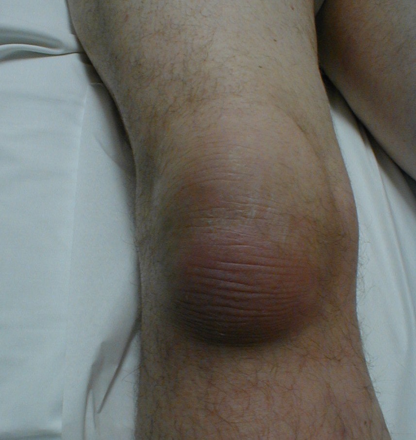
The patient presents with pain in the anterior aspect of the knee and swelling on the lower side of the patella. The swelling is distal to the swelling of prepatellar bursitis though it becomes difficult to differentiate in cases of large swellings. There might be a history of frequent kneeling.

Extremes of flexion and extension would cause pain. There is tenderness in the area of swelling.

If the swelling is anterior to the patellar tendon, it is superficial infrapatellar bursitis. If the swelling is posterior to the patellar tendon, deep infrapatellar bursitis is presented. Both can be differentiated on MRI.

Lab Studies

CBC, ESR, CRP may be needed if the infection is suspected. Anitnuclear antibody, rheumatoid factor, and anti – citric citrullinated peptide tests should be done in cases of autoimmune diseases.

Aspiration and analysis of bursal fluid should be done to rule out infectious or rheumatic causes. Fluid is tested for biochemical and cytological analysis. Gram stain and culture for infection can be done in cases of infection.

Imaging

Xrays are normal in most of the cases but are done to identify any underlying bony pathology. Tendon calcifications may be found in chronic bursitis.

Ultrasonography may help in injection application or aspirations in deep bursae.

MRI is generally not done in routine but is able to define the lesion better and even differentiate from abscess and tumors.

MRI is able to differentiate between superficial and deep infrapatellar bursitis.

Treatment of Infrapatellar Bursitis

The treatment includes rest, ice compression and elevation [RICE therapy]. Anti-inflammatory drugs like NSAIDs decrease pain and swelling by reducing inflammation. In large swellings, bursa is aspirated and intrabursal steroid injections could be used.

The cause is identified and repeated trauma is avoided [e.g. frequent kneeling]

In infectious cases, antibiotics are used, first broad spectrum and the culture based antibiotic should be given.

Most of the infections can be treated by antibiotic therapy but some cases would require surgical drainage as well.

In chronic or recurring cases, removal of bursa may be sought.


Spread the Knowledge
  • 1
    Share
  •  
    1
    Share
  • 1
  •  
  •  
  •  
  •  

Filed Under: Knee

About Dr Arun Pal Singh

Arun Pal Singh is an orthopedic and trauma surgeon, founder and chief editor of this website. He works in Kanwar Bone and Spine Clinic, Dasuya, Hoshiarpur, Punjab.

This website is an effort to educate and support people and medical personnel on orthopedic issues and musculoskeletal health.

You can follow him on Facebook, Linkedin and Twitter

Reader Interactions

Leave a Reply Cancel reply

Your email address will not be published. Required fields are marked *

This site uses Akismet to reduce spam. Learn how your comment data is processed.

Primary Sidebar

muscle power grading

Muscle Power Grading

Muscle power grading is a frequent and important part of the neurological examination. The neurological examination consists of sensory and motor examination. Muscle power grading is part of motor system examination. Before muscle power grading is done, limbs or trunk are examined for the tone of the muscles, the bulk of the muscles. The limbs […]

wound-dressings

Wound Dressings and Coverings

Wound care dressings and procedures aim at rapid covering healing of skin defects are for better wound healing. The best method for healing a wound is to close the defect by surgical means as quickly after the injury as possible. However, this may not always be possible. Where primary closure is not possible [ large […]

osgood schlatter disease

Osgood Schlatter Disease Causes Symptoms and Treatment

Osgood Schlatter disease is osteochondrosis of tibial tubercle that results from repetitive quadriceps contraction through the patellar tendon at its insertion on tibial tubercle. This occurs in the period before puberty as the tibial tubercle is growing and is susceptible to strain. It is is a self-limiting disorder of the knee. It is named after […]

Wrist sign in Marfan Syndrome

What Is Marfan Syndrome?

Marfan syndrome is an inherited connective-tissue disorder transmitted as an autosomal dominant trait which has features like tall stature, ectopia lentis, mitral valve prolapse, aortic root dilatation, and aortic dissection. Pathophysiology Marfan syndrome results from mutations in the fibrillin-1  gene on chromosome 15, which encodes for the glycoprotein fibrillin. Fibrillin is used as a major […]

bicipital tendonitis relevant anatomy

Bicipital Tendonitis or Biceps Tendonitis

Bicipital tendonitis [also called biceps tendinitis or tendionpathy], or, is an inflammatory process of the long head of the biceps tendon. Bicipital tendinitis can occur due to impingement or rotator cuff injuries, tears of glenoid labrum, and other intra-articular pathologies. Pathophysiology of Bicipital Tendonitis Biceps has its origins from two heads. The long head biceps […]

Classification of scaphoid fractures

Scaphoid Fractures – Classification, Presentation and Treatment

The scaphoid is most frequently fractured carpal bone. Scaphoid fractures account for up to 15% of acute wrist injuries and 50% to 80% of carpal fractures. Scaphoid fractures have been reported in people aged 10-70 years but commonly occur in young, active individuals,  peak incidence being in the second and third decades of life. Scaphoid […]

Spinal cord Injury Levels

Spinal Cord Injury Levels

Spinal cord injury levels can be expressed in many ways and it is important to understand the correlations. This has an important bearing in marking the level of injury and correlating with the anatomy. For example, if an injury occurs at T10-11 vertebral level, it is going to damage her lower thoracic as well as […]

Browse Articles

Footer

Pages

  • About
    • Policies
    • Contact Us

Featured Article

Patellar Instability – Causes, Presentation and Treatment

Patellar instability or Patellofemoral instability is a frequent cause of symptoms of anterior knee pain and episodes of mechanical instability.It … [Read More...] about Patellar Instability – Causes, Presentation and Treatment

Search Articles

© Copyright: BoneAndSpine.com