Last Updated on July 31, 2019
Kohler disease is osteochondrosis of the navicular bone, first described by Kohler in 1908. In this condition also called navicular osteochondrosis, there is avascular necrosis navicular bone.
Alban Kohler was a German radiologist who first described this condition.
Exact occurrence of Kohler disease is not known. The disorder is more frequent in children aged 5-10 years but can begin as early as 2 years. Boys are more commonly affected than girls but girls are reported to be affected at a younger age. This happens because the onset of ossification in girls occurs earlier by six months than boys.
Causes of Kohler Disease
Cause of Kohler disease is not known.
The navicular bone is the last tarsal bone to ossify in children. Ossification centers of the navicular appear between the ages of 1.5 and 2 years in girls and 2.5 and 3 years in boys.
It is thought that this unossified bone may get compressed between the already ossified talus and the cuneiforms when the child gets becomes heavier. Compression of the bone and the vessels involves the vessels leads to ischemia causing clinical symptoms. This, in due course, is followed by revascularization by branches from the perichondral ring of vessels and thus making a prognosis of this lesion is always excellent.
Main blood supply of navicular comes from dorsalis pedis artery. It crosses the dorsal surface of the navicular bone and gives off three to five branches. Some small branches also come from the medial plantar artery and supply the plantar surface. A network of vessels is created around the bone and come from the perichondrium toward the center of the cartilage. Sometimes only a single dorsal or plantar vessel is found.
Presentation of Kohler Disease
Painful limp is the most common presentation. The child may walk with an increased weight on the lateral side of the foot. There may be local tenderness of the medial aspect of the foot over the navicular bone. The child can walk with an increased.
Swelling and soft tissue redness may be present.
Lab Studies
Not required
Imaging Studies

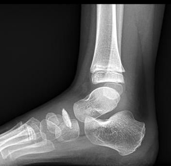
Routine radiographs may show navicular as flattened, fragmented and thinned like a wafer. It may show patchy sclerosis and often soft tissue swelling is also present.
CT and MRI are usually not required but may be necessary if pain persists 6 months after casting or the diagnosis is not clear or to the tarsal coalition.
Treatment of Kohler Disease
Standard treatment is weight bearing below-the-knee I slight varus [heel turned inward] and equinus for 6-8 weeks. Equinus and varus relax navicular from the strain from the posterior tibialis strain.
Following cast removal, arch supports are given for about 6 months.
If the symptoms and condition are very mild, soft arch supports may be the sole mode of treatment.
Most of the treated patients get relieved by 3 months. However. In untreated patients, symptoms may be present for 15 months.
If the pain still persists after 6 weeks period of casting, a new cast is applied for 6 supplementary weeks. Other causes of foot pain, like talar coalition or an accessory navicular, may also be investigated.
Prognosis
Nearly all the patients recover with excellent function. Patients treated with cast immobilization had a quicker resolution of symptoms than patients treated without casting. .Rarely navicular becomes distorted and sclerotic and, the head of the talus becomes flattened and the joint between two becomes fibrillated, and degenerative changes like osteophyte formation may occur. Such patients with persistent pain need arthrodesis of the talonavicular joint. The calcaneocuboid joint is included because most of its role is lost when the talonavicular joint is fused.