Last Updated on October 27, 2023
Lumbar spine anatomy is unique in being strong and flexible. Lumbar spine provides mobility in many different planes including flexion, extension, side bending, and rotation, and at the very same time is strong enough to support body weight and the load of different postures. The lumbar spine protects the highly sensitive spinal cord and spinal nerve roots.
Lumbar spine consists of 5 vertebrae linked by joint capsules, flexible ligaments/tendons, and muscles.
Lumbar spine consists of 5 lumbar vertebrae named L1 to L5. The topmost lumbar vertebra is designated as L1 and it articulates with the D12 vertebra of the dorsal spine. The lowermost lumbar vertebra is L5 which articulates with S1 of the sacrum.
 The five vertebral bodies and intervertebral discs of the lumbar spine withstand significant physiologic loads. The intervertebral segment of the lumbar spine consists of a three-articulation complex, the disc-vertebral body, and two posterior apophyseal (facet) joints [superior and inferior].
Structural Arrangement of Lumbar Spine
The vertebral bodies are a cylindrical mass of cancellous bone with a cortical shell. In between are the discs consisting of the annulus fibrosus, the nucleus pulposus, and the cartilaginous and bony endplates of the vertebral bodies.
The vertebral bodies and discs from the anterior column of the spine, which is responsible for resisting approximately 80% of axial compressive loads and maintaining spinal rigidity and alignment.
The intervertebral disc maintains separation of the vertebral bodies.
The anterior part of the disk is thicker than the posterior part and is responsible for most of the lumbar lordosis because the vertebral bodies are near uniform in shape.
Nearly two-thirds of lumbar lordosis is localized between the L4 and S1 segments.
The posterior column of the spine consists of
- Spinous processes
- Lamina
- Transverse processes
- Facet Joints
These structures control movement and resist forces.
The facet joints are hyaline joints formed by the superior and inferior articular processes of subjacent vertebrae with a fibrous joint capsule.
The superior articular process faces posterior and medial, and the inferior articular process faces lateral and anterior.
The joint surface is oblique [The facet joints are oriented 120 degrees to 150 degrees] to the sagittal plane [Plane that divides the body into right and left halves]. It facilitates slight flexion and extension or sagittal plane rotation as each articular process slides on the other.
This orientation resists anterior or posterior translation in normal anatomic alignment and axial rotation in the horizontal plane.
Lumbarization and Sacralization
These are anatomical variations of the lumbar spine
In lumbarization, the first sacral vertebra, which is normally fused to rest of sacrum remains unfused. On x-ray, the lumbar spine appears to have six vertebrae and the sacrum appears to have only four segments instead of five.
In sacralization, the last lumbar vertebra (L5) fuses to the sacrum on one side or both, or to the ilium, or giving an appearance of four lumbar vertebrae.
These anomalies are observed at about 3% of people and most are asymptomatic.
Read more on lumbarization and sacralization.
Vertebral Canal and Its Content
The vertebral canal contains the spinal cord [which terminates at L2] its meninges, spinal nerve roots, and blood vessels supplying the cord and other structures.
Bony Lumbar Spine Anatomy
Lumbar Vertebrae

L2, L3, and L4 are typical lumbar vertebrae. L1 and L5 have some peculiarities and are atypical lumbar vertebrae.
Vertebrae are composed of the following 3 functional parts:
- The vertebral body
- The vertebral (neural) arch
- The bony processes (spinous and transverse
Body
The vertebral body of each lumbar vertebra is large, wider from side to side than from front to back, and a little thicker in front than in back. It is flattened or slightly concave above and below, concave behind, and deeply constricted in front and at the sides.
There are no rib facets present, a feature that distinguishes lumbar vertebrae from thoracic.
Vertebral bodies are together connected by the intervertebral discs. The size of the vertebral body increases from L1 to L5.
L5 vertebra has the heaviest body, smallest spinous process, and thickest transverse process.
The epiphyseal ring is the cortical ring on the distal surface of a vertebra. It acts as an anchor to annulus fibrosus of disc and also serves as growth zone.
The surface within epiphyseal ring is covered by hyaline cartilage.
Vertebral or Neural Arch
All things posterior to the vertebral body form the vertebral arch.
Each vertebral arch is composed of
- 2 pedicles
- 2 laminae
- 7 bony processes held together by facet joints and ligaments
- 1 spinous process
- 4 articular processes – 2 superior and 2 inferior
- 2 transverse processes
Pedicles are posteriorly directed strong bony bars from the upper part of the vertebral body which meets laminae to complete neural arch posteriorly.
Pedicle sagittal width increases from L1 to L5 [9 mm to up to 18 mm] and increases in angulation as well in the axial plane [10-20 degrees]
The laminae form the posterior portion of the vertebral arch. In the upper lumbar region the lamina is taller than wide but in the lower lumbar vertebra, the lamina is wider than tall. The lamina connects the spinous process to the pedicles.
Neural arch along with the posterior surface of vertebra form vertebral foramen which harbors nerve root in meninges.
Bony Processes
The spinous process is thick, broad, and projects backward. Its posterior end is rough and uneven.
Four articular processes arise from each vertebra, two projecting upward and two downward from the junctions of pedicles and laminae.
The facets on the superior processes are concave, and look backward and medialward. Those on the inferior are convex and are directed forward and lateralward.
Superior articular processes form a facet joint with the inferior articular process of the superior vertebra. Similarly, inferior articular processes form a facet joint with superior articular facets of the inferior vertebra.
The transverse processes project in the coronal plane. These long and slender processes are horizontal in the upper three lumbar vertebrae and incline a little upward in the lower two.
These processes from the junctions of the pedicles and laminae in upper three vertebrae but in the lower two they move forward and originates from the pedicles and posterior parts of the vertebral bodies.
Three tubercles are seen in the transverse process of a lower lumbar vertebrae
- Lateral or costiform process [lateral]
- Mammillary process [superior]
- Accessory process [inferior]
Neural Foramina
Neural foramina are openings in the vertebral column on either side from where nerve roots exit.
They are numbered by vertebra above them. For example, the L1 foramen is beneath L1 vertebrae.
Each foramen is bounded superiorly and inferiorly by the pedicle, anteriorly by the intervertebral disc and vertebral body, and posteriorly by facet joints. The same numbered spinal nerve root, recurrent meningeal nerves, and radicular blood vessels pass through each foramen.
Fifth Lumbar Vertebrae
The fifth lumbar vertebra has a body much deeper in front than behind to accommodate the prominence of the sacrovertebral articulation.
The spinous process is smaller and there is a wide interval between the inferior articular processes.
Transverse processes are thicker and originate anteriorly than usual.
Intervertebral Discs
Discs connect and cushion the vertebrae while allowing enough movement.
Their size varies depending on the adjacent vertebrae size.
Each disc consists of the nucleus pulposus which is a mucoid substance embedded with reticular and collagenous fibers, surrounded by the annulus fibrosus, a fibrocartilaginous lamina.
The anterior fibers of the annulus fibrosus are strengthened by the anterior longitudinal ligament. Posteriorly most of the fibers are attached to the cartilage plate.
[Read on Disc anatomy and Biomechannics]
Vertebral joints
There are symphyseal joints between the vertebral bodies. These are formed by a layer of hyaline cartilage on each vertebral body and an intervertebral disc between the layers.
Facet joints or zygapophysial joints or Z-joints are the joints between inferior and superior articular processes of vertebrae as discussed earlier.
Facet joints are synovial joints and permit simple gliding movements.
The region between the superior articular process and the lamina is the pars interarticularis. This area is important as it is areas of spondylolisthesis.
Ligaments of Lumbar Spine
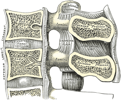
Anterior Longitudinal Ligament
The anterior longitudinal ligament runs down the anterior surface of the vertebral bodies from the occiput to the sacrum. The anterior longitudinal ligament is thicker and narrower in the thoracic region. The ligament firmly attaches to the edges of the vertebral bodies.
This is thick and slightly more narrow over the vertebral bodies and thinner but slightly wider over the intervertebral discs. It also loosely attaches to the annulus of the disc.
It is a three-layered ligament – superficial, intermediate and deep.
Posterior Longitudinal Ligament
The posterior longitudinal ligament arises from the posterior aspect of the basiocciput in the skull, is continuous with the membrana tectoria, and runs over the posterior surfaces of the bodies of the vertebrae, down to the coccyx. It is within the vertebral canal.

Intertransverse Ligaments
These ligaments are situated between the transverse processes. In the thoracic region, they are closely connected with the deep muscles of the back.
Capsular Ligaments
The capsular ligaments are attached to the articular margins of the articular processes. The fibers are oriented perpendicular to the facet joint and are stronger in the thoracic and lumbar region than in the cervical region.
Ligamentum Flavum
Also called Yellow Ligament, the ligamentum flavum connects the anteroinferior edge of the lamina to the posterosuperior edge of the lamina below. The ligamentum flavum is thicker in the thoracic region. This ligament is composed mainly of elastic fibers, and that elasticity serves to preserve the upright posture and to assist the vertebral column to resume after flexion.
Interspinous Ligaments
The interspinous ligaments connect adjacent spinous processes and runs obliquely from the anterior inferior aspect of the spinous process above to the posterior superior aspect of the spinous process
Supraspinous Ligament
This strong ligament is a strong fibrous cord, which connects together the tips of the spinous processes from the C7 to the sacrum.
Iliolumbar Ligament
The iliolumbar ligaments attach to the ilium and L5 vertebra transverse processes act to limit anterior translation and rotation making the lumbosacral junction a very stable articulation.
Muscles of Lumbar Spine

Image Credit: Medscape
Functionwise, the muscles of the lumbar spine can be grouped together as extensors, flexors, lateral flexors, and rotators.
Lumbar spine musculature works in a synergistic way from both the left and right side muscle groups.
Extensor Muscles
Erector Spinae
Erector spinae [sacrospinalis] is the primary extensor and largest group of intrinsic back muscles. In the lower lumbar spine, the erector spinae appears as a single muscle but divides into 3 vertical columns of muscles (iliocostalis, longissimus, spinalis) in the upper part. Iliocostalis is most lateral and spinalis is most medial.
All of these have a common origin from a thick tendon attached to the sacrum, the lumbar spinous processes, and the iliac crest.
Transversospinal
This muscle group lies deep to the erector spinae and takes origin from the mamillary processes, from the laminar area just medial to the posterior sacral foramina in the sacrum, from the tendinous origins on the erector spinae, and the medial surface of the posterior superior iliac spine.
These include
- 3 semispinalis muscles, spanning 4-6 vertebral segments
- Semispinalis thoracis
- Semispinalis cervicis
- Semispinalis capitis
- Multifidus, spanning 2-4 vertebral segments
- Rotatores, spanning 1-2 vertebral segments
- Rotatores cervicis
- Rotatores thoracis
- Rotatores lumborum
- Interspinales
- Intertransversarii
It is a fasciculated muscle group and each fascicle is directed superomedially toward the inferior and medial margin of the lamina and adjacent spinous process. The superficial layer attaches from 3-4 levels above, the intermediate layer attaches 2 levels above, and the deep layer attaches 1 level above.
Along with extension, this muscle group also takes part in the rotation.
Segmental
These form the deepest layer. These can be divided into two groups. The levatores costarum are not present in the lumbar spine but the other group formed by interspinales and intertransversarii is.
Interspinales is between the spinous processes of contiguous vertebrae. Intertransversarii pass between adjacent transverse processes. They are postural stabilizers and work to increase the efficiency of larger muscles.
Forward flexors
Iliothoracic
These are abdominal wall muscles namely rectus abdominis, external abdominal oblique, internal abdominal obliquus, and the transversus abdominis. These are also called extrinsic flexors.
Femorospinal
Psoas major and iliacus muscles form this group of iliac flexors.
Lateral flexors
This movement is a combination of side bending and rotation. The muscles that participate in side bending are ipsilateral the oblique and transversus abdominal muscles and quadratus lumborum.
Rotators
Lumbar spine rotation is by oblique muscles such as the extensors and lateral flexors assisted by the antagonist muscle groups to counter the primary effect of these muscles.
The transversospinal muscle group, when contracted unilaterally cause the trunk to rotate in the contralateral direction. They are divided into 3 groups: muscles. The rotatores lumborum are small, irregular, and variable muscles connecting the superoposterior part of the transverse process of the vertebra below to the inferolateral border of the lamina of the vertebra above.
Blood Supply of Lumbar Spine
Paired lumbar arteries arise from the aorta, opposite the bodies of L1-L4. Each pair travels anterolaterally around the side of the vertebral body and gives off various branched opposite when reaches laterally to the vertebral canal.
Periosteal and equatorial branches supply the vertebral bodies
Spinal branches enter the intervertebral foramen, divide into smaller anterior and posterior branches to supply vertebral body, vertebral arch, meninges, and spinal cord.
The larger branches of the spinal branches continue as radicular or segmental medullary arteries and supply nerve roots and to the spinal cord, respectively.
Venous plexuses are formed by veins along the vertebral column both inside and outside the vertebral canal. Vasivertebral veins emerge from the foramen on the posterior surfaces of the vertebral bodies and drain into the internal vertebral venous plexuses. The intervertebral veins anastomose with veins from the cord and venous plexuses and drain into the lumbar segmental veins.