Last Updated on September 12, 2019
Mallet finger is a finger deformity caused by disruption of the extensor tendon mechanism distal to the distal interphalangeal joint caused by a bony or tendon injury
It is a common injury and occurs as a workplace injury or in sports. It commonly occurs in young to middle-aged males. It is also seen in older females.
It is the most common closed tendon injury seen in athletes. Mallet finger is also called a drop, hammer, or baseball finger.
Long, ring and small fingers of dominant hand are most commonly affected by this injury.
Relevant Anatomy
The injury that causes mallet finger disrupts the tendinous attachment of the extensor mechanism.

The terminal extensor tendon is an extension of the dorsal mechanism or extensor mechanism which refers to the unique arrangement of extensor tendon, interossei, and lumbricals. The dorsal mechanism flexes the metacarpophalangeal joints and extends the proximal and distal joints.
The terminal extensor tendon is approximately 1 mm thick and 4 mm wide. It crosses the distal interphalangeal joint in the midline and inserts onto the dorsal lip of the distal phalanx, proximal to the germinal nail matrix.
It causes the extension of the distal interphalangeal joint.
Thus tendon avulsion [with or without bone chip] or laceration of tendon results in active extension power of the DIP joint is lost. The unopposed force of flexor tendons brings the joint in an abnormally flexed position.
Causes, Mechanism of Injury and Pathophysiology of Mallet Finger
The injury could occur as traumatic blow or laceration.
The traumatic blow causes suddenly forced flexion of the tip [distal interphalangeal joint] of the extended finger.
This is a classic mechanism of mallet finger injury where the finger is in rigid extension and the force causes flexion of the distal phalanx.
The cause of the force could a ball striking while playing such as volleyball or basketball.
A finger that gets stuck and flexed such as in a bedspread or slipcover can also result in mallet finger.
Another interesting mechanism is pushing off a sock with extended fingers.
Fracture of the dorsal articular surface of the distal phalanx may cause a mallet finger as well. These are noted as bony avulsions on x-ray. The size of the avulsion may vary from a fleck of bone to fragment involving more than third of the articular surface.
Another mechanism is a sharp or crushing laceration to the dorsal aspect of the finger at about distal interphalangeal joint.
With the loss of extensor force at the distal interphalangeal joint, the entire power of extension is directed to the PIP joint. Over a period, it may result in the hyperextension of the proximal interphalangeal joint. Together, PIP extension and DIP flexion give rise to a swan-neck deformity.
Doyle’s Classification of Mallet Finger Injuries
- Type I
- Closed injury with or without small dorsal avulsion fracture
- Type II
- Open injury (laceration)
- Type III
- Open injury (deep soft tissue abrasion involving loss skin and tendon substance)
- Type IV
- Mallet fracture
- A = distal phalanx physeal injury (children)
- B = fracture fragment involving 20% to 50% of articular surface (adult)
- C = fracture fragment >50% of articular surface (adult)
- Mallet fracture
Clinical Presentation of Mallet Fingers
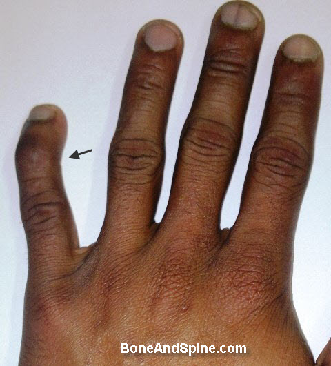
After an injury, often caused by a typical mechanism as discussed above, the patient complains of inability to extend the distal joint actively. Pain may not be there. In fact, the majority of the cases are painless.
The examination will reveal that dorsum of the finger is tender and swollen.
The examination will show slightly flexed finger at the distal inerphalangeal joint.
In late cases, hyperextension of the proximal interphalangeal may result in swan neck deformity.
Differential Diagnoses
- Jammed Finger
- Phalangeal Fractures
- Swan neck deformity in Rheumatoid Arthritis
Imaging
Posteroanterior and lateral x-rays of the affected finger are required.
The x-rays can differentiate between bony mallet injury and a tendinous injury. Any associated fracture or dislocation may be found.
The x-ray should be centered on the distal interphalangeal joint. X-ray of the whole hand is not deeded unless there is injury to other parts of the hand as well.
Treatment of Mallet Finger
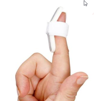
Most of the mallet fingers are treatable by nonoperative methods even the bony avulsions. Conservative treatment includes splinting the finger in extension [not hyperextension]. Once extension splinting has been initiated, it should be maintained without even a momentary lapse for the prescribed treatment period.
The splint is to be worn 24 hours, daily. Tendinous injuries require splint for 6-8 weeks and bony injuries for 4-5 weeks. the splint may be required longer in old injuries. Splinting may be extended for 2-4 weeks if extensor lag is noted after removal.
The splints could be customized for the patient or one of the commercially available pre-molded plastic splints could be used.
After the desired period and in absence of extensor lag, the splints are weaned over the next 1-2 weeks. The splint should be used at night and with activities that put the joint at risk.
Surgical treatment is required less commonly.
In the case of a bony fragment, the surgery consists of either closed reduction and percutaneous pinning or open reduction and internal fixation. The indications are
- Absolute indications
- volar subluxation of distal phalanx
- Relative indications
- More than 50% of the articular surface involved
- More than 2mm articular gap
The fracture is fixed by K-wire.
Some patients who cannot wear splints for the required 6-8 weeks for vocational reasons, may be given an internal splintage by Kwire. With a digital block, a 0.035-in. K-wire can be inserted across the joint to serve as a temporary internal splint.
In case of tendinous injury, surgical reconstruction of the terminal tendon is the surgery of choice in cases of chronic injury (more than 12 weeks) with the healthy joint. This procedure has a high rate of complications.
Other procedures which are rarely done are arthrodesis of distal interphalangeal joint for painful, arthritic joint, and swan neck deformity correction
The procedures employed are
- Direct repair/tendon advancement
- Tenodermodesis
- Spiral oblique retinacular ligament reconstruction
Complication
- Extensor lag
- Usually slight extensor lag present but there is no functional deficit
- Swan neck deformities
- Skin necrosis due to splint
- Mostly affects dorsum of the finger
- Seen when the splint is applied in hyperextension
- Complications after surgery
- Infection
- Hardware problems
- Stiffness of distal interphalangeal joint
Prognosis
A mallet finger which is not treated does not affect function mostly.
With treatment, a functional and cosmetic normal finger can be obtained.
Swan neck deformity when present can produce disability.
References
- Alla SR, Deal ND, Dempsey IJ. Current concepts: mallet finger. Hand N Y. 2014 Jun. 9(2):138-44
- Valdes K, Naughton N, Algar L. Conservative treatment of mallet finger: A systematic review. J Hand Ther. 2015 Jul-Sep. 28 (3):237-46.