• Skip to primary navigation
  • Skip to main content
  • Skip to primary sidebar
  • Home
  • TeleConsult
  • About
  • Newsletter/Updates
  • Contact Us
  • Policies

Bone and Spine

Orthopedic health, conditions and treatment

  • General Ortho
  • Procedures
  • Spine
  • Upper Limb
  • Lower Limb
  • Pain
  • Trauma
  • Tumors

Microfracture Chondroplasty

By Dr Arun Pal Singh

In this article
    • Indications for Microfracture Chondroplasty
    • Timing of Procedure
    • Contraindications
    • Surgical Technique of Microfracture Chondroplasty
      • Rehabilitation
      • Related

The microfracture chondroplasty is a technique to treat full-thickness cartilage defects that lead to the formation of new joint surface cartilage.

It includes the debridement of the damaged area and puncturing the underlying bone with a micro-instrument or pick to allow bleeding from the bone. The resulting blood clot that fills the cartilage defect has specialized cells which have the potential to transform into new cartilage.

This procedure is reserved for more significant areas of joint surface damage where the lesion is full thickness and down to the underlying bone. Microfracture can be performed on any size lesion, but better results have been obtained with lesions smaller than 400 mm.

The articular cartilage of the knee is 2 to 4 mm thick, avascular tissue devoid of nerves and lymphatics. Articular cartilage defects that extend for the full thickness to subchondral bone rarely heal without intervention. These injuries could be due to acute trauma or repetitive strain.

Initially developed for knee defects, microfracture chondroplasty is used now for hip, talus, elbow, and shoulder as well.

Indications for Microfracture Chondroplasty

  • Full-thickness articular cartilage defects in the weight-bearing area
  • Unstable cartilage flaps in these regions that extend to the subchondral bone

Timing of Procedure

Acute full thickness cartilage loss is treated as soon as possible.

Chronic lesions or degenerative lesions are first treated conservatively initially for at least 12 weeks by activity modification, physical therapy, NSAIDs, and joint injections, as appropriate.

Patients who have failed conservative treatment for chronic or degenerative lesions then become candidates for microfracture chondroplasty.

Contraindications

  • Patients who unable or unlikely to complete the rehabilitation
  • Advanced age
  • Generalized degenerative changes
  • Inflammatory arthritis
  • Unstable knees are relative contraindications.

Most of these are relative contraindications

Surgical Technique of Microfracture Chondroplasty

The procedure is done under either regional or general anesthesia.

The patient is kept supine on a standard operating table.

Standard arthroscopic portals can be used for microfracture.

The lesion is identified for microfracture and probed thoroughly to make sure that all bordering cartilage is stable.

A creation of stable full-thickness borders of cartilage surrounding a central lesion is optimal for microfracture chondroplasty. It provides some degree of protection to the regenerating tissue that is forming in the treated lesion.

Using a shaver, all loose flaps of cartilage are removed.

Then proceed to remove the calcified cartilage layer by using a hand-held curette.

microfracture-chondroplasty
Image Credit: Orthop Clin N Am 36 (2005) 419 – 426

This will complete the bed preparation.

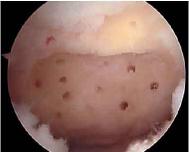
Now advance the arthroscopic awls through the subchondral bone to create multiple small holes or microfractures to a depth that allows the return of fat droplets or blood from the cancellous bone.

The usual depth of the holes is 2 to 4 mm.

Keep the holes as close together as possible without breaking one into the next.

The microfracture awl produces a roughened surface in the subchondral bone. This rough surface allows the marrow clot to adhere more easily.

Once the microfracture is complete, the pieces of equipment is removed from the joint. Do not use intra-articular drains. All portal sites are closed with suture and wounds are dressed.

Rehabilitation

Put the patient on the continuous passive motion, as tolerated. Stationary biking without resistance and deep-water exercise programs are started at 1 to 2 weeks after microfracture.

Patients with weight-bearing lesions are also prescribed toe-touch weight bearing with crutches for 6 to 8 weeks.

Patients who have undergone microfracture chondroplasty in the patellofemoral joint are treated in a locking brace with flexion limited to 20 degrees.

This is done to eliminate shear forces across the lesion during walking. Brace is removed/unlocked only for continuous passive motion exercises. The brace is worn for at least 8 weeks. The brace is then discontinued after the full range of motion is gradually achieved over approximately 1 week.

Anti-inflammatory medications and physical therapy modalities are prescribed as appropriate.

Related

Spread the Knowledge
8
Shares
 
8
Shares
 8   

Filed Under: Procedures

About Dr Arun Pal Singh

Arun Pal Singh is an orthopedic and trauma surgeon, founder and chief editor of this website. He works in Kanwar Bone and Spine Clinic, Dasuya, Hoshiarpur, Punjab.

This website is an effort to educate and support people and medical personnel on orthopedic issues and musculoskeletal health.

You can follow him on Facebook, Linkedin and Twitter

Primary Sidebar

Browse Articles

bone island xray

Bone Island or Enostosis

A bone island or enostosis is a common benign bone lesion. It is asymptomatic and does not require treatment. It is usually seen as an incidental finding on x-ray, computed tomogram and MRI. A bone island is a focus of mature cortical bone within the cancellous bone.  Pelvis, femur, and other long bones are the […]

Child on Denis Browne Splint

Congenital Talipes Equinovarus or Club Foot

Congenital talipes equinovarus or club foot or CTEV as it is often known as is a congenital deformity affecting single foot or both. Club foot may result from a bony, a muscular or a neuropathic error, or maybe termed idiopathic. Idiopathic club foot is the most frequent and unless specified otherwise, the term congenital talipes […]

ganglion cyst on wrist

Ganglion Cyst Presentation and Treatment

Ganglion cyst is the most common benign tumor of the hand. It is thought to be degeneration of the mucoid connective tissue. It most commonly occurs on the dorsal wrist in 60% cases and is seen as bump on the wrist.This arises generally from scapholunate joint but can also arise from the radioscaphoid or scaphotrapezial […]

intrathecal pain pump

Intrathecal Pain Pump Concept, Use and Risks

An intrathecal pain pump is a surgically implanted drug delivery system that sends pain medication directly to the spinal cord. This unique treatment option may be recommended to selected patients of chronic pain who have not experienced adequate pain relief from conservative approaches. The intrathecal pain pump is a small battery-powered, programmable pump which is […]

Olecranon Bursitis

Olecranon Bursitis Presentation and Treatment

Olecranon bursitis is inflammation of olecranon bursa which is a subcutaneous bursa overlying the olecranon process of the ulna. Bursa functions to reduce the friction between olecranon and skin. Olecranon bursitis may result from inflammation caused by various factors like repetitive trauma and infection of the bursa. Normally, olecranon bursa is Superficial locations of the […]

Fracture Head Second Metacarpal With Fracture Shaft of First Metacarpal Fixred With Kwire and Finger Distractor

Metacarpal Fractures Causes, Presentation and Treatment

Metacarpal fractures are fractures of long tubular bones of hand that connect carpal bones to proximal phalanges. Metacarpal fractures account for 30-40% of all hand fractures. The metacarpal neck is the most commonly fractured region which constitutes around 10% of all hand fractures. They mostly occur between age group 10-30 years. Men are affected more. […]

Foot Sprain

Sprain Causes and Treatment

A sprain is an injury that occurs to ligaments causing stretching or tearing of ligaments. Ligaments are fibrous structures that join two bones across the joints. For example, ankle ligaments connect tibia and fibula bones to the talus and calcaneum across the ankle joint. Sprain originates from French word espriandre which means to wring. Therefore […]

© Copyright: BoneAndSpine.com
Manage Cookie Consent
The site uses cookies. Please accept cookies for a better visiting experience.
Functional Always active
The technical storage or access is strictly necessary for the legitimate purpose of enabling the use of a specific service explicitly requested by the subscriber or user, or for the sole purpose of carrying out the transmission of a communication over an electronic communications network.
Preferences
The technical storage or access is necessary for the legitimate purpose of storing preferences that are not requested by the subscriber or user.
Statistics
The technical storage or access that is used exclusively for statistical purposes. The technical storage or access that is used exclusively for anonymous statistical purposes. Without a subpoena, voluntary compliance on the part of your Internet Service Provider, or additional records from a third party, information stored or retrieved for this purpose alone cannot usually be used to identify you.
Marketing
The technical storage or access is required to create user profiles to send advertising, or to track the user on a website or across several websites for similar marketing purposes.
Manage options Manage services Manage vendors Read more about these purposes
View preferences
{title} {title} {title}
 

Loading Comments...