• Skip to main content
  • Skip to primary sidebar
  • Skip to footer
  • General Ortho
  • Procedures
  • Spine
  • Upper Limb
  • Lower Limb
  • Pain
  • Trauma
  • Tumors
  • Newsletter/Updates
  • About Us
  • Contact Us

Bone and Spine

Orthopedic health, conditions and treatment

Motion Preservation Surgery of Spine

By Dr Arun Pal Singh

In this article
    • Motion Preservation Surgeries for Cervical Spine
      • Cervical Total Disc Replacement
    • Motion Preservation Surgeries for Lumbar  Spine
      • Anterior Motion Preservation Devices
      • Posterior Motion Preservation Devices
      • None of the devices have withstood the test of time, and most are still in various stages of investigation.
    • Kyphoplasty
    • References

Motion preservation surgery in the spine refers to a group of surgeries which aim at preservation of the spine motion by replicating normal or near normal biomechanics. The fundamental to all these surgeries is the use of devices, that replicate these functions.

Decompression and fusion are classic procedures of spine surgery. Motion preservation surgeries are relatively newer concept.  The intent of motion preservation surgeries is to provide a better treatment alternative that preserves the function of the spine well.

Motion preservation surgery of the spine is a complex, constantly evolving field and the research is ongoing.

Motion Preservation Surgeries for Cervical Spine

Cervical motion preservation surgery mainly consists of total disc replacement.

Cervical Total Disc Replacement

In cervical total disc replacement, there is the removal of the diseased disc and implantation of the arthroplasty device. The goal is neural decompression, restoration of near normal motion so as to minimize adjacent level disease.

Two devices for commercial use are the ProDisc-C (Synthes Spine USA) and Prestige ST (Medtronic, Memphis, TN).

Clinical trials are underway to determine clinical effectiveness versus anterior cervical compression and fusion

Complications of cervical total disc replacement include fracture, subsidence, migration of the implant, heterotopic ossification, and hardware failure or loosening. Subsidence, as in lumbar TDR, is a gradual process whereby the device settles into the adjacent vertebral body.

Motion Preservation Surgeries for Lumbar  Spine

In the lumbar spine, motion preservation devices can be loosely grouped into anterior and posterior motion-preserving devices. anterior motion preservation devices

Anterior Motion Preservation Devices

These devices are intended to replicate the normal motion of the disc space.

Lumbar total disc replacement

Lumbar TDR devices vary widely in shape and composition. Following  devices are in use

  • Charite device (DePuy Spine)
  • FlexiCore device (Stryker Spine)
  • ProDisc-L device (Synthes Spine)
  • NuVasive’s XL (Nuvasive)

Most of these devices require an invasive anterior approach. NuVasive’s XL is implanted indirectly through a far-lateral approach.

charite disc and xray. One of the motion preservation surgeries
Charite artificial disc model and x-rays after implantation
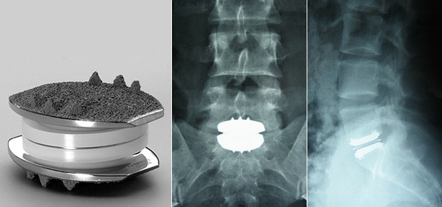
Image Credit: bnasurg

Complications associated with lumbar total disc replacement are hemorrhage, vascular injury, infection, peritoneal entry, vertebral fracture. Late complications of  total disc replacement are subsidence of implant, implant migration, heterotopic ossification, adjacent-level degenerative disc disease

The clinical effectiveness has not been unequivocally determined.

Disc Nucleus Replacement

In partial disc replacement, the diseased nucleus pulposus is replaced or augmented with an injectable or preformed device. Annulus fibrosis is not replaced. The devices are in development

[Read anatomy of disc]

Posterior Motion Preservation Devices

These devices address pain originating from the posterior elements from the facet joints, ligaments, tendons, or muscles. Posterior motion preservation devices are also beneficial in spinal stenosis.

These devices can be put into three general categories:

Interspinous Process Spacers

These devices are designed to distract (open) the central canal and foramen[opening where the nerve roots come out of spinal canal]. These devices are designed to address pain and activity restrictions from spinal stenosis. Some of them may be used to treat degenerative disc disease.

The interspinous devices may be implanted as a day surgery procedure under sedation or under light anesthesia, thereby decreasing the risk of extensive open surgery in elderly.

Various interspinous spacer devices in use are X-STOP, Wallis interspinous device, and DIAM Spinal Stabilization System.

Posterior Dynamic Stabilization Devices

This acts as an internal brace for the spine. Posterior dynamic stabilization devices allow controlled motion in such as way as to achieve more normal movement of the spine. These devices are typically used to treat patients with spondylolisthesis and degenerative disc disease. As of now, these devices have been approved for use with fusion but not for independent use as dynamic stabilizers.

Facet Replacement or Total Element Replacement Devices

These are used in facet pain or lumbar spinal stenosis as in many patients, spinal stenosis is due to degeneration of the facet joints. Facet replacement devices replace the facet joints in the back of the spine to limit or control motion, and total element replacement devices replace all the elements in the back of the spine.

None of the devices have withstood the test of time, and most are still in various stages of investigation.

Kyphoplasty

Kyphoplasty is a minimally invasive treatment developed to treat vertebral compression fractures in the spine. Compression fractures cause back pain and make daily activities like walking and lifting difficult. The principle of the kyphoplasty is to strengthen the spine and possibly eliminate pain.

Read more about kyphoplasty

References

  • J. Errico. Why a mechanical disc? Spine J, 4 (Suppl 6) (2004), pp. 151S–157S
  • Yin, X. Yu, S. Zhou, et al. Is cervical disc arthroplasty superior to fusion for treatment of symptomatic cervical disc disease? A meta-analysis. Clin Orthop Relat Res, 471 (2013), pp. 1904–1919
  • Wei, Y. Song, L. Sun, et al.     Comparison of artificial total disc replacement versus fusion for lumbar degenerative disc disease: a meta-analysis of randomized controlled trials.   Int Orthop, 37 (2013), pp. 1315–1325.
  • Yajun, Z. Yue, H. Hiuxin. A meta-analysis of artificial total disc replacement versus fusion for lumbar degenerative disc disease.   Eur Spine J, 19 (2010), pp. 1250–1261
  • C. Wang, P.M. Arnold, J.T. Hermsmeyer, et al. Do lumbar motion preserving devices reduce the risk of adjacent segment pathology compared with fusion surgery?  Spine, 37 (22S) (2012), pp. S133–S143
  • E. Ziegler, J. Glenn, R.B. Delamarter. Five-year adjacent-level degenerative changes in patients with single-level disease treated using lumbar total disc replacement with ProDisc-L versus circumferential fusion.  J Neurosurg Spine, 17 (6) (2012), pp. 504–511


Spread the Knowledge
  • 1
    Share
  •  
    1
    Share
  • 1
  •  
  •  
  •  
  •  

Filed Under: Spine

About Dr Arun Pal Singh

Arun Pal Singh is an orthopedic and trauma surgeon, founder and chief editor of this website. He works in Kanwar Bone and Spine Clinic, Dasuya, Hoshiarpur, Punjab.

This website is an effort to educate and support people and medical personnel on orthopedic issues and musculoskeletal health.

You can follow him on Facebook, Linkedin and Twitter

Reader Interactions

Comments

  1. shobhit mehta says

    May 17, 2016 at 11:18 pm

    Is this technique being used by big hospitals like AIIMS ?

  2. Dr Arun Pal Singh says

    May 18, 2016 at 9:52 am

    These are newer methods and not all of them have been incorporated in clinical practice. However, few surgeons with special interest may be practising them. If you wish to know, you would need to ask the concerned surgeon rather than looking for hospital.
    Take care.

  3. auri says

    July 3, 2016 at 12:39 pm

    Hi Doc I’m really scared as I’m due for surgery soon I had an mri done after I fell of a roller chair at work results were annular tear on l4 and l5 with dosc protrusio and disc dessication my surgeon had advised me that it needs to be removed as the disc is black due to all the fluid leaking out I have been in soo much pain for the past 12 months awaiting the procedure and the time is near.i was told here is not other natural options as If I don’t do the op I could end up paralysed because the div is now sitting on my spinal cord.is there any advice I’m really worried that after surgery I my not walk I don’t know how advanced South Africa is in terms of spine procedures.

  4. Dr Arun Pal Singh says

    July 19, 2016 at 5:48 pm

    Sorry if this reply does not get you in time.
    Auri, what you need is a second opinion after taking a personal consultation with another doctor. Only after examination and evaluation it can be told whether you need surgical procedure or not.

  5. auri says

    December 9, 2016 at 12:41 am

    Hi Doc how would I know if motion preservation surgery is available in South Africa I have a herniated disc on l4 and l5 and disc dessication so it’s black and has protruded into the spinal canal I suffer pain daily and medication is not so strong anymore

  6. Dr Arun Pal Singh says

    December 11, 2016 at 10:26 pm

    Auri,

    Sorry! I am not aware of this in South Africa.

Leave a Reply Cancel reply

Your email address will not be published. Required fields are marked *

This site uses Akismet to reduce spam. Learn how your comment data is processed.

Primary Sidebar

Acromioclalvicular injuries

Acromioclavicular Joint Injuries and Their Treatment

Acromioclavicular joint injuries are also known as shoulder separations, acromioclavicular joint separation or acromioclavicular joint dislocation and occur as a result of downward force on the acromion. Acromioclavicular joint injuries occur most commonly in sporting activities. These are most commonly seen in young adults although an increasing trend is noted in children due to increased […]

Dysplasia Epiphysealis Punctata lethal

Dysplasia Epiphysealis Punctata

Dysplasia epiphysealis punctata is a rare condition characterized by the presence of stippled epiphyses at birth in association with other connective tissue disturbances. It is also known under a variety of names such as chondrodystrophia foetalis calcificans, stippled epiphyses, or congenital multiple epiphyseal dysplasia. It is also called Conradi Disease. Two main forms are recognized […]

Essex Lopresti Fracture

Essex Lopresti Fracture

The Essex Lopresti fracture is also called longitudinal radioulnar dissociation and consists of fracture of the radial head, rupture of the interosseous membrane, and disruption of the distal radioulnar joint. It is important to recognize  and have high index of suspicion because Essex Lopresti fracture is often missed. Isolated radial head fracture can be successfully […]

Comartment syndeome-complications of fractures

What are Complications of Fractures

Fractures treatment in modern times allows most of the fractures to heal without a problem. But complications of do occur sometimes which may affect management and outcome. Complications of fractures can be acute or chronic. Acute complications of fractures are mostly due to initial injury and generally as a result of the initial trauma and […]

Harms of prolonged sitting

Prolonged Sitting, Its Harmful Effects and Remedies

Prolonged sitting occupies most of our daily time both at home, office or transport. Human bodies are made for moving around as that is what we did for thousands of years. But rapidly developing technology has led to a sedentary life. Modernization of the offices and shifting of emphasis from paper to computer has also […]

Flexor Muscles of Hand and Wrist

Forearm Muscles – Anatomy and Function

The forearm is the area of upper extremity between the elbow and wrist. The bony structure of the forearm is formed by two bones – radius and ulna. There are twenty forearm muscles which are arranged in two compartments – anterior and posterior. The anterior compartment contains flexor muscles and is also called the flexor […]

Carpal Tunnel Diagram

Scaphoid Bone Anatomy

The scaphoid bone is one of the carpal bones of the wrist on the lateral side of the wrist. Carpal bones are arranged in two rows and scaphoid is most lateral of the proximal row. The carpus is made up of 8 carpal bones, which are arranged in two rows. Proximal row It contains (from […]

Browse Articles

Footer

Pages

  • About
    • Policies
    • Contact Us

Featured Article

Scapular Dyskinesis – Causes, Tests and Treatment

Scapular dyskinesis is a term that denotes loss of control of normal scapular motion, physiology or mechanics. It is also called sometimes as scapular … [Read More...] about Scapular Dyskinesis – Causes, Tests and Treatment

Search Articles

© Copyright: BoneAndSpine.com