Last Updated on October 29, 2023
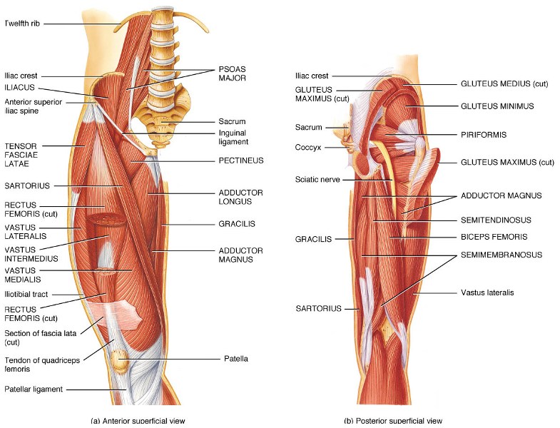
Muscles of the hip joint are those muscles that cause movement in the hip. The muscles of the hip consist of four main groups.
Gluteal Group
The gluteal muscles cover the lateral surface of the ilium and include
- Gluteus maximus
- Gluteus medius
- Gluteus minimus
- Tensor fasciae latae
Adductor group
This group includes
- Adductor brevis
- Adductor longus
- Adductor magnus
- Pectineus
- Gracilis
The adductors all originate on the pubis and insert on the medial, posterior surface of the femur, with the exception of the gracilis which inserts just below the medial condyle of the tibia.
Iliopsoas group
The iliacus and psoas major comprise the iliopsoas group.
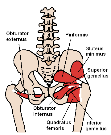
Lateral rotator group
This group consists of
- Obutrator externus
- Obturator internus
- Piriformis
- Superior and inferior gemelli
- Quadratus femoris.
These six originate at or below the acetabulum of the ilium and insert on or near the greater trochanter of the femur.
Other hip muscles
Rectus femoris and the sartorius can cause some movement in the hip joint but these muscles primarily move the knee, and not generally classified as muscles of the hip.
The hamstring muscles, which originates mostly from the ischial tuberosity and insert on the tibia/fibula, also assist with hip extension.
Gluteus Maximus
Origin
Gluteus maximus is a large muscle of the hip that arises from
- Posterior gluteal line of the inner upper ilium
- Rough portion of bone including the crest, immediately above and behind it
- Posterior surface of the lower part of the sacrum and the side of the coccyx,
- Lumbodorsal fascia
- Sacrotuberous ligament
- Fascia covering the gluteus medius.
Insertion
The fibers are directed obliquely downward and lateralward. The gluteus maximus has two insertions – iliotibial band of the fascia lata and gluteal tuberosity between the vastus lateralis and Adductor Magnus.

Action
- Hips extension
- External rotation of the hip
- Upper fibers assist in adduction
- Lower fibers assist in abduction
Gluteus Medius

The gluteus medius extends from the ilium to the femur and acts as abductor and medial rotator of the hip.
Origin
The outer surface of ilium just below the crest
Insertion
Greater trochanter
Actions
- The abduction of the hip
- Anterior fibers – Internal rotation and flexion
- Posterior fibers – External rotation and extension
Gluteus Minimus
The gluteus minimus lies beneath the gluteus medius and is its companion in attachments and functions.
Both muscles lie beneath the superficial and largest of the posterior thigh muscles, the gluteus maximus.
Origin
Gluteus Minimus originates from the outer surface of the ilium beneath the gluteus medius
Insertion
Greater trochanter of the femur
Actions
- The abduction of the hip
- Internal rotation
- Flexion of the hip
Tensor Fascia Lata
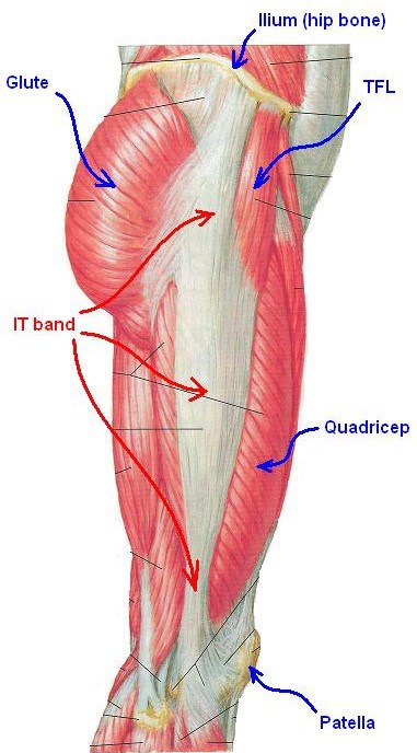
The tensor fasciae latae is a tensor of the fascia lata, a fibrous sheath in the thigh.
Origin
It arises from the anterior part of the outer lip of the iliac crest, anterior superior iliac spine, and part of the outer border of the notch below it and from the deep surface of the fascia lata.
Insertion
It is inserted between the two layers of the iliotibial band of the fascia lata about the junction of the middle and upper thirds of the thigh.
Actions
The tensor fasciae latae tautens the iliotibial band and braces the knee, especially when the opposite foot is lifted. It aids in the following movements
- Flexion of the hip
- Internal rotation
- The abduction of the hip
Adductor Brevis
Origin
It originates by a narrow origin from the outer surfaces of the superior and inferior rami of the pubis, between the gracilis and obturator externus.
Insertion
Its fibers, passing backward, lateralward, and downward, are inserted, by an aponeurosis, into the line leading from the lesser trochanter to the linea aspera and into the upper part of the linea aspera, immediately behind the pectineus and upper part of the adductor longus.
Actions
- Adduction
- External rotation
- Flexion (weak)

Adductor Longus
The adductor longus lies ventrally on the adductor magnus, and near the femur, the adductor brevis is interposed between these two muscles.
Origin
It originates from the front of the pubis just below its crest
Insertion
It is inserted into the middle third of the medial lip of the linea aspera.
Nerve Supply
Anterior division (sometimes the posterior division) of the obturator nerve.
Actions
- Adduction of hip
- Flexion (weak)
Adductor Magnus
The adductor magnus is a large triangular muscle that is located posterior to the adductor longus
Origin
The muscle consists of two parts.
- Adductor part [pubofemoral portion, adductor portion, or adductor minimus] – Arises from the ischiopubic ramus
- Extensor Part [ischiocondylar portion, n, or “hamstring portion] – Arises from the tuberosity of the ischium
The ischiocondylar portion of the adductor magnus is considered a muscle of the posterior compartment of the thigh [due to its common embryonic origin, innervation, and action similar to the hamstring group of muscles] while the pubofemoral portion of the adductor magnus is considered a muscle of the medial compartment.
Insertion
Adductor part
Fibers which arise from the ramus of the pubis are short, horizontal in direction, and are inserted into the rough line of the femur leading from the greater trochanter to the linea aspera, medial to the gluteus maximus.
Fibers from the ramus of the ischium are directed downward and laterally with different degrees of obliquity, to be inserted, by means of a broad aponeurosis, into the linea aspera.
Ischiocondylar Part
Fibers arising from the tuberosity of the ischium, form a thick fleshy mass which descends almost vertically and end about the lower third of the thigh in a rounded tendon.
This tendon is inserted into the adductor tubercle on the medial condyle of the femur and is connected by a fibrous expansion to the line leading upward from the tubercle to the linea aspera.
Nerve Supply
- Adductor portion is innervated by the posterior division of the obturator nerve.
- Hamstring portion is innervated by the tibial nerve.
Actions
- Adduction of hip
- External rotation of the hip
- Extension of hip
Pectineus
It is a flat, quadrangular muscle situated at anteromedial aspect of the thigh. The pectineus muscle is the most anterior adductor of the hip. ]
Origin
The muscle arises from
- Pectineal line of the pubis
- Slight extent from the surface of the bone in front of it between the iliopectineal eminence and pubic tubercle
- From the fascia covering the anterior surface of the muscle
Insertion
The fibers pass downward, backward, and lateral, to be inserted into the pectineal line of the femur which leads from the lesser trochanter to the linea aspera.
Innervation
Femoral nerve (L2 and L3) + Accessory obturator nerve [in 20% people].
Actions
Hip flexion.

Gracilis
Gracilis is a thin flattened muscle on the medial side of the thigh. It is most superficial muscle on medial side of thigh thin and flattened, broad above, narrow and tapering below.
Origin
Gracilis arises by a thin aponeurosis from the anterior margins of the lower half of the symphysis pubis and the upper half of the pubic arch.
Insertion
Muscle fibers run vertically downward, end in a rounded tendon which passes behind the medial condyle of the femur and curves around the medial condyle of the tibia where it flattens, and inserts into the upper part of the medial surface, below the condyle immediately above that of the semitendinosus muscle.
Its upper edge is overlapped by the tendon of the sartorius muscle and together they form the pes anserinus. The pes anserinus is separated from the medial collateral ligament of the knee-joint by a bursa.
A few of the fibers of the lower part of the tendon are prolonged into the deep fascia of the leg.
Nerve Supply
Anterior division of obturator nerve (L2, L3)
Actions
- Adduction at the hip
- Internal rotation of leg at the knee
- Flexion (weak)
Obturator Externus
The external obturator muscle is short external rotator muscle of hip joint. It is a flat, triangular muscle on the anterior wall of the pelvis.
Origin
It arises from two-thirds of the outer surface of the obturator membrane, the inferior pubic ramus, and the ramus of the ischium.
Insertion
The fibers converge and pass posterolateral and upward, to form a tendon that runs across the back of the neck of the and is inserted into the trochanteric fossa of the femur.
Nerve Supply
Posterior branch of obturator nerve
Actions
The external obturator muscle acts as the lateral rotator of the hip joint. As a short muscle around the hip joint, it stabilizes the hip joint as a postural muscle.
Obturator Internus
Origin
Obturator internus originates from the internal surface of obturator membrane and posterior bony margins of the obturator foramen.
Insertion
It inserts on the medial surface of greater trochanter of the femur, in common with superior and inferior gemelli.
Nerve Supply
Nerve to obturator internus (L5, S1, S2)
Actions
It is a short external rotator of hip joint.
Gemelli
The superior gemellus muscle arises from the ischial tuberosity and inserts on the greater trochanter. It is an external rotator of the thigh. It is supplied by the nerve to obturator internus
The inferior gemellus muscle arises from the ischial tuberosity and inserts on the medial surface of the greater trochanter of the femur and rotates the thigh outward. It is supplied by the nerve to Quadratus femoris.
Both are external rotators of hip.
Piriformis
Piriformis is one of six short external rotators [others being superior gemellus, inferior gemellus, obturator internus, obturator externus, and quadratus femoris muscles – together known as deep gluteals] and participates in the abduction of hip also.
The piriformis muscle is found deep to the gluteus maximus and inferior to the gluteus minimus. Its name is because of its a pear-like shape.
Origin
It takes origin along the anterior region of the sacrum.
Insertion
It extends laterally, passing through the greater sciatic foramen and crossing the hip joint to insert on the greater trochanter of the femur.
Nerve Supply
Nerve to the piriformis (L5, S1, and S2)
Actions
External rotation of hip
Quadratus Femoris
The quadratus femoris is a flat, quadrilateral skeletal muscle located on the posterior side of the hip joint.
Origin
It takes origin from ischial tuberosity
Insertion
It inserts into intertrochanteric crest.
Nerve Supply
Nerve to quadratus femoris
Actions
It is a strong external rotator and adductor of the thigh.
Iliopsoas
The iliacus and psoas major muscles are closely related in structure and function and are often referred to as a single muscle, the iliopsoas.
These muscles are distinct in the abdomen, but usually indistinguishable in the thigh.

Origin
The psoas major originates along the lateral surfaces of the vertebral bodies of T12 and L1-L3 and their associated intervertebral discs. The iliacus originates in the iliac fossa of the pelvis.
Insertion
The psoas major unites with the iliacus at the level of the inguinal ligament and crosses the hip joint to insert on the lesser trochanter of the femur. The iliopsoas is involved in flexion and lateral rotation (supination) of the thigh. If the limb is fixed they involve in flexion of the trunk.
Innervation
The psoas major is innervated by direct branches of the anterior rami of the lumbar plexus ( L1-L3) while the iliacus is innervated by the femoral nerve (L2-L4).
Actions
Iliopsoas are the powerful flexors of the thigh at the hip joint. The iliopsoas is important for standing, walking, and running.
Sartorius

Sartorius, the longest muscle in the human body is a long, thin, band-like muscle.
Origin
The sartorius muscle arises by tendinous fibers from the anterior superior iliac spine, runs obliquely across the upper and anterior part of the thigh in an inferomedial direction.
Insertion
It descends as far as the medial side of the knee, passes behind the medial condyle of the femur to end in a tendon which curves anteriorly to join the tendons of the gracilis and semitendinosus muscles to form the pes anserinus, and inserts into the medial surface of proximal tibia.
Nerve supply
Anterior (or superficial) branch of the femoral nerve
Actions
The sartorius muscle functions as flexor, abductor and external rotator of the hip joint. At the knee joint the sartorius helps to flex the leg. Sitting cross-legged, making ankles rest on opposite knee are the movements where sartorius muscle is used.
Turning the foot to look at the sole demonstrates all four actions of the sartorius.
Rectus Femoris
The rectus femoris constitutes quadriceps muscle along with vastus lateralis, vastus medialis, and vastus intermedius.
Origin
Rectus femoris arises by two tendons
- one, the anterior or straight, from the anterior inferior iliac spine
- The other, the posterior or reflected, from a groove above the rim of the acetabulum.
Insertion
The two heads unite and spread into an aponeurosis which is prolonged downward on the anterior surface of the muscle, and from this, the muscular fibers arise. The muscle gradually narrows into a flattened tendon and is inserted into the base of the patella.
Nerve Supply
Femoral Nerve
Actions
For the hip, the rectus femoris acts as flexor. It is also strong extensor of the knee as part of quadriceps muscle of knee.
Hamstring Muscles
Semimembranosus, semitendinosus and biceps femoris are three hamstring muscles that originate above the hip joint and insert below the knee joint.
Semitendinosus and semimembranosus extend the hip when the trunk is fixed and flex the knee. They also medially rotate the lower leg when the knee is bent.
The long head of the biceps femoris extends the hip as when beginning to walk. Both short and long heads flex the knee and laterally rotates the lower leg when the knee is bent.