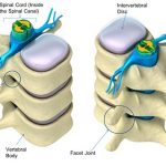
Vertebral canal or spinal canal is the long tubular space in the vertebral column formed by contiguous placement of vertebral foramen through which the spinal cord passes along with other contents Vertebral foramina are stacked on one another to form a long canal. In the intervertebral spaces, the canal is protected by the ligamentum flavum […]
Anatomy
Flexor Tendon Pulley System of Hand
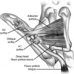
Flexor tendon pulley system consists of annular ligaments of the fingers or A pulleys, and cruciate pulleys [C pulleys]. Flexor pulley system consists of following Palmar Aponeurosis pulley 5 Annular pulleys 3 Cruciform pulleys. Together, these form a fibro-osseous tunnel on the palmar aspect of the hand through which passes the deep and superficial flexor […]
Bone Anatomy and Physiology
The adult human skeleton has a total of 206 bones, excluding the sesamoid bones. The appendicular skeleton has 126 bones, axial skeleton 74 bones, and auditory ossicles six bones. [Read more about different bones and their classification] Each bone possesses a unique shape. But the bones across the body do possess similarities in terms of […]
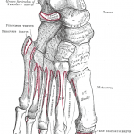
Anatomy of Metatarsal Bones and Phalanges
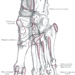
Metatarsal Bones or Metatarsus There are five metatarsal bones in the foot which are numbered from medial to lateral side. Common Features of Metatarsal Bones Each metatarsal bone is a miniature long bone and has following parts Base Base is the proximal end of metatarsal bone and articulates with tarsal bones. It is set obliquely […]
Tarsal Bones Anatomy
There are seven tarsal bones. The proximal row is formed by the talus above, and the calcaneus below. The distal row contains, from medial to lateral side, medial cuneiform, the intermediate cuneiform, the lateral cuneiform and the cuboid. Navicular is interposed between the talus and the three cuneiform bones. In other words, it is interposed […]
Ankle Joint Anatomy
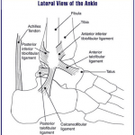
The ankle joint is a hinged synovial joint. In an absolute sense, its primary movements are moving the foot up and down, called dorsiflexion and plantar flexion respectively. However, this joint works in tandem with subtalar joints [talocalcaneal and talocalcaneonavicular], thus adding to the range of motion of this ankle-subtalar joint complex. Anatomically subtalar (talocalcaneal) […]
Calcaneus Anatomy and Attachments
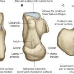
Calcaneus is bone of the hindfoot and is the largest tarsal bone. Calcaneus forms the prominence of the heel. Structure of Calcaneus Calcaneus is roughly cuboidal in shape. It is directed forwards, upwards and laterally. It has six surfaces. Anterior Surface The anterior surface is the smallest surface of the bone. It is covered by […]
Talus Bone Anatomy
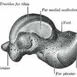
Talus is a tarsal bone of hindfoot. There are about 26 bones in the human foot grouped into 3 parts – tarsal bones, metatarsal bones, and phalanges. The foot itself can be divided into 3 parts Hindfoot or rearfoot Midfoot Forefoot Hindfoot is formed by talus and calcaneus, two of the seven tarsal bones. Rest […]
Tibiofibular Joint Anatomy
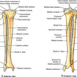
Tibiofibular joints are articulations between tibia and fibula. Superior tibiofibular joint is articulation between head of fibula and upper tibia. At ankle, the articulation between tibia and fibula is called inferior tibiofibular joint. Two bones are connected by interosseous membrane which is also sometimes called middle tibiofibular joint. Superior Tibiofibular Joint The superior tibiofibular joint […]
Meniscus Anatomy, Function and Significance
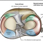
Knee menisci, two in number, are made of fibrocartilaginous tissue and present between lateral and medial condylar articulation of tibia and femur. Knee meniscus or simply meniscus refers to either the lateral or medial fibrocartilaginous structure. The menisci are also known as semi-lunar cartilages because of their crescent shape. Not very long ago, menisci were thought […]