Last Updated on March 3, 2024
Osteochondrosis is a self-limiting developmental derangement of normal bone growth, primarily involving the centers of ossification in the epiphysis. It usually begins in childhood as a degenerative or necrotic condition. By definition, osteochondrosis is an aseptic ischemic necrosis.
In broad terms, osteochondroses are a group of unrelated lesions that mostly affect immature skeletons [children], and involve either epiphysis or apophysis and bone in the apophyseal region. Epiphyseal ossification centers are primarily affected. It has been suggested to be a type of ischemic necrosis.
As the research on osteochondrosis is gathering more data, a few osteochondroses are labeled as a phenomenon rather than a disease or syndrome.
Most of the osteochondroses are more common in males. However, Freiberg disease and juvenile osteochondritis dissecans of the elbow involving the capitellum (seen often in javelin throwers) is common among females.
It mostly is seen in ages between 10-15 years. The condition is very rare in people younger than 10 years and older than 50.
The condition mostly affects people between 10 and 15 years. They are very rare in people under 10 and over 50 years.
Epiphysis, Physis and Apophysis
In an adult skeleton, the bone consists of metaphyses and diaphysis. However, an immature skeleton contains physeal and epiphyseal regions that are present at the ends beyond the metaphyseal region. After the bone has attained its maximum growth potential, the physis, epiphysis, and metaphysis are joined.
Epiphysis
The epiphysis is the end part of the bone and is separated from the metaphysis by the physis or growth plate. The epiphyseal part contributes to the joint formation.
Physis
Physis is also called a growth plate and is a thin cartilage that separates epiphysis from metaphysis.
Apophysis
The apophysis is a secondary ossification center that is located in the non-weight-bearing part of the bone [appears separated from bone] but eventually fuses with it over time It is a site of tendon or ligament attachment. It is also called traction epiphysis.

Types of Osteochondrosis
It divides the osteochondroses into articular, nonarticular, and physeal types.
Articular or Epiphyseal osteochondrosis
This type involves articular regions and could be either primary involvement of the articular and epiphyseal cartilage (Freiberg disease that involves second toe) or secondary involvement of the articular and epiphyseal– (Perthes disease of hip, Kohler disease of foot) In case of secondary involvement, the primary pathology is the ischemic necrosis of the bone.
Nonarticular osteochondrosis
These can occur in any part of the skeleton. These can be at the attachment of tendons (Osgood-Schlatter’s disease of the knee and Monde-Felix disease) or ligamentous attachments like the vertebral ring, and impact sites like Sever disease that is also associated with sports and overuse.
Physeal osteochondrosis
• Long bones – Tibia vara (Blount disease)
• Scheuermann disease
Another type of classification of osteochondrosis is juvenile which occurs in childhood, and adult which occurs in adults.
Another set of terms used about osteochondrosis is osteochondritis deformans or “osteochondritis dissecans depending on the site and its presumed cause.
Osteochondritis deformans affects the primary ossification center in young children (Legg-Calve-Perthes disease, Kohler’s disease, Panner’s disease) and is thought due to vascular causes whereas osteochondritis dissecans affects bone and cartilage of weight-bearing areas in older children such as femoral condyle, talus etc and is thought to be of traumatic origin. But the cause and effect is neither that straightforward nor established as of now.
A List of eponyms of osteochondrosis is given below
Location |
Eponym |
| Foot | |
| Talus | Mouchet or Diaz disease |
| Navicular (Tarsal scaphoid) | Kohler’s disease |
| Calcaneal apophysis | Sever disease or phenomenon |
| Accessory tarsal navicular or os tibiale externum | Haglund disease |
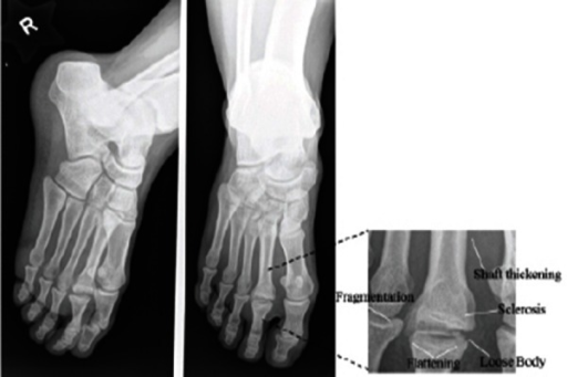
| Second metatarsal | Freiberg disease |
| Fifth metatarsal base | Iselin disease |
| Medial cuneiform | Bushcke disease |
| Spine and Pelvis | |
| Vertebral epiphysis | Scheuermann kyphosis |
| Iliac crest | Buchman disease |
| Symphysis pubis | Pierson disease |
| Ischiopubic junction | Van Neck disease or phenomenon |
| Ischial tuberosity | Valtancoli disease |
| Vertebral body | Legg-Calve-Perthes Disease |
| Lower Limb | |
| Patella | Kohler disease |
| Capital epiphysis of the femur | Legg-Calve-Perthes disease |
| Greater trochanter of the femur | Mandl or Buchman disease |
| Distal tibial epiphysis | Lewin disease |
| Proximal tibial epiphysis | Blount disease |
| Tuberosity of the tibia | Osgood-Schlatter disease |
| Secondary patellar center | Sinding-Larsen-Johansson syndrome |
| Lesser trochanter of the femur | Monde-Felix disease |
| Upper Limb | |
| Lunate | Kienboch’s disease |
| Phalanges | Thiemann syndrome |
| Metacarpal heads | Mauclaire disease |
| Proximal epiphysis of the radius | Schaefer disease |
| Carpal scaphoid | Preiser disease |
| Distal epiphysis of the ulna | Burns disease |
| Medial humeral condyle | Froelich disease |
| Lateral humeral condyle | Froelich disease |
| Capitellum of the humerus | Panner disease |
| Humeral head | Hass disease |
| Clavicle | Friedrich disease |
Pathophysiology
Most osteochondroses occur shortly after the bony nucleus appears, around the middle of the growth spurt. Physeal and apophyseal osteochondrosis is often caused by injury/trauma in the form of repeated sports injuries or overuse.
For example,physeal osteochondrosis is often found in gymnasts or tennis players in the distal radius.
In the case of apophyses, the growth of bones and the myotendinous units attached may be at a different rate owing to increased traction forces, especially during rapid growth.
However, most of these injuries are reversible early on if the repetitive action causing the injury is stopped.
There is no clear agreement on the cause of epiphyseal osteochondrosis. There are many theories like endocrine, genetic, vascular, and traumatic theories
The initial event whether due to trauma or vascular cause is suggested as ischemic necrosis of the ossification center. This leads to degeneration of the epiphyseal osseous nucleus . The interference with blood supply and failure of the bony centrum to enlarge may lead to this resulting in disordered proliferation of the cartilaginous cells.
Secondary changes like fragmentation, collapse, and sequestrum formation may occur but would vary with the affected region.
Apart from trauma and vascular causes, the following causes have been suggested to play a role
- Thrombosis/ embolism
- Trace element deficiency (copper, zinc)
- Infection
- Exposure to passive smoking
Clinical Presentation and Diagnosis
On examination localized swelling, tenderness, and limited movement of a nearby joint may be noted. Lower limb involvement may show gait problems.
Growth disturbance and secondary deformities may occur in the late stages.
The presentation of osteochondrosis depend on the site of involvement and on the stage of progression. Several patients may not have symptoms. Pain localized to the affected site is the usual presentation in the initial stages. The pain might worsen as the condition worsens. For example, pain on kneeling in cases of Osgood-Schlatter disease. Some conditions like Perthes disease may present with a referred pain.
There may or may not be a history of overuse.
The pain may progress as follows
- Pain after physical activity only
- Pain on physical activity without affecting function
- Pain during and after physical activity and affected function
- Pain even without activity or minimal activity
The pain may not be localized in epiphyseal and physeal osteochondrosis but in cases of apophyseal osteochondrosis, the pain is well localized.
On examination, the following may be seen in some cases
- Tenderness
- Swelling over the site
- Nearby joint movement restriction
- Limp-in lower limb pathologies
Some conditions may show growth changes and deformity. For example
- Blount disease- shortening and tibia vara
- Scheuermann disease -Kyphosis
- Perthes disease- Shortening and hip changes
Any presence of systemic signs like fever or myalgia in other parts should be noted.
Lab Investigations
Laboratory findings are mostly normal.
However, it is important to rule out other disorders, and following tests would help when suggested.
• Hemogram
• Erythrocyte sedimentation rate (ESR)
• C-reactive protein (CRP) level
• Serum calcium
• Serum phosphate
• Alkaline phosphatase (ALP) levels.
Other lab tests as deemed necessary on an individual basis may be done.
Imaging
X-rays
X-rays in the early stage show an epiphysis of reduced size with increased opacity. The structural irregularities if visible can be seen. As the disease advances, cartilage fissures and fibrillation may occur. Irregular bony trabeculae may be visible. X-rays done in the revascularization phase show osteoporosis, physis deformation, and underlying bone support defects.
The revascularization phase results in osteoporosis, absorption of necrotic tissue, and deformation. Incomplete restoration of the blood supply and poor protection can lead to misshaped epiphysis. Associated changes may be seen in adjoining metaphyseal areas in these cases. Joint arthritis sets in late cases.
A few of the known deformities associated with different osteochondrosis are-
- Mushroom-shaped femoral head or joint incongruity- Perthes disease
- Kyphosis – Scheuermann disease
- Cubitus valgus – Panner disease
- Madelung deformity in radial epiphysis
- Genu recurvatum and patella alta in Osgood-Schlatter disease
MRI
MRI can pick up the disease at the asymptomatic stage and before x-ray changes. However, it is required in a few cases especially when differentiation from inflammatory conditions and infection is required.
Bone Scan
Bone scintigraphy is again a sensitive and specific test that can pick up the problem before symptoms. The bone scan can indicate the phase of the disease as well and thus can be used as a prognostic marker.
Treatment
Most of the cases of osteochondrosis are self-limiting. Medical and supportive therapy constitute the mainstay of treatment.
The goal is to obtain a congruous, mobile, and painless joint.•
The part is protected from additional trauma and stress. This facilitates ossification and prevention of deformity. Any loose osteochondral fragments need to be removed.
Surgery is indicated only in cases where conservative therapy has failed or when it would help to reduce late disability. Deformity correction surgeries may be done in late stages if required.
Medical Treatment
- Rest to joint
- Ice application
- Drugs for controlling pain and inflammation – ibuprofen, diclofenac etc.
- Traction– provides pain relief, counter spasms and prevents deformity. Needed in selected cases
- Immobilization – by plaster or brace
- Assisted weight bearing when required
- Physical therapy
Surgical Treatment
Surgical treatment is usually undertaken when conservative methods are not effective. Surgery is also used when it may help restore the reparative process as to improve outcomes over conservative means.
Arthroscopic procedures may be done to facilitate regeneration by microfracture technique or to pin or fix a salvageable fragment. Osteotomy is done to correct the deformity.
Salvage surgeries and reconstructive procedures are done in to improve situations in cases with poor outcomes. These may include arthrodesis, arthroplasty, or bone lengthening procedures where required.
The surgery needs to be individualized.
Prognosis
Osteochondrosis is mostly a self-limiting disorder, the outcome is usually good. However, those uncommon cases who don’t respond to treatment either conservative or surgical are associated with poor prognosis.
References
- West EY, Jaramillo D. Imaging of osteochondrosis. Pediatr Radiol. 2019 Nov. 49 (12):1610-1616. [Link]
- Atanda A Jr, Shah SA, O’Brien K. Osteochondrosis: common causes of pain in growing bones. Am Fam Physician. 2011 Feb 1;83(3):285-91.
- Achar S, Yamanaka J. Apophysitis and Osteochondrosis: Common Causes of Pain in Growing Bones. Am Fam Physician. 2019 May 15;99(10):610-618. [Link]