• Skip to primary navigation
  • Skip to main content
  • Skip to primary sidebar
bone and spine logo

Bone and Spine

Your Trusted Resource for Orthopedic Health Information

  • Home
  • About
  • Contact Us
  • Policies
  • Show Search
Hide Search
You are here: Home / Basics and Biomechanics / Pars interarticularis Anatomy and Significance

Pars interarticularis Anatomy and Significance

Dr Arun Pal Singh ·

Last Updated on October 28, 2023

Contents hide
1 What is Pars Interarticularis?
2 Significance of Pars Interarticularis
2.1 Scottie Dog Sign and Collar Sign

What is Pars Interarticularis?

The Pars Interarticularis is a special region of the lamina between the superior and inferior articular processes. The term literally means part between the articulations in Latin language and is kind of bony bridge that joins these two upper and lower facets.

pars interarticularisThus one vertebral has two pars interarticularis.

Thus facet joints above are connected to the joints below in one vertebra through the pars interarticularis.

An isthmus is a narrow portion of land that joins two larger bodies of land. Pars interarticularis is considered isthmus between two facet joints.

Significance of Pars Interarticularis

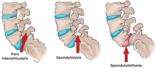
Pars interarticularis may develop a stress fracture in young athletes. Defect in the pars could lead to spondylolisthesis

When we can see on x-ray in the lateral view, vertebra, L4. L5 and S1  face downward and forward as they form the lower end of the normal lordotic curve.

spondylolysis and spondylolisthesis
Spondylolysis and spondylolisthesis. Image credit: Open i

These two vertebrae bear the entire weight of the spine above.

The inferior facets prevent the sliding of lower vertebra forward by acting as a buttress. Thus L5 inferior facets act as a buttress to S1 slide and L4 inferior facets to L5 slide.

An analogy could be given that of door stops. Pars interarticularis joins these “door stops” to the upper structures of the vertebra(the pedicles).

This puts these structures under quite a significant load, especially under the impact.

Sports, jumping, and activities involving hyperextension of the spine [as in gymnasts] can cause overloading.

This could lead to stress fractures of pars interarticularis.

While stress fracture can heal with rest, the continued activity could lead to complete fracture and then nonunion which then becomes a cause for a forward slip of vertebra [no more doorstop effect]. The disc is not able to resist this as dis has poor shear strength.

The disc, under stress, stretches and tears paving a way for the unresisted forward slip.

The fractures normally occur on pars of both sides but occasionally can only on one side. A unilateral pars fracture causes the other, intact pars to bear all the stress and this eventually leads to fracture of that pars too.

Before the conclusion, I would like to mention a similar sounding term – spondylolysis. It means there is a break or fracture of the pars but no slip.

When the slip occurs, the term used is spondylolisthesis.

Pars fracture or spondylolisthesis could be discovered as an incidental finding when a child is evaluated for some other reason or could present as back pain that is aggravated by continued lumbar loading.

The diagnosis has to be radiologically confirmed though clinically it could be suspected in some cases.

Scottie Dog Sign and Collar Sign

Scotty dog sign on oblique view spine, shows intact pars interarticularis
Case courtesy of A.Prof Frank Gaillard, Radiopaedia.org. From the case rID: 7552

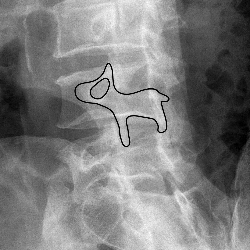
This sign suggests the normal appearance of the lumbar spine seen on oblique view x-ray. Following elements contribute to formation of Scottie dog with:

  • Transverse process forms the nose
  • Pedicle forms the eye
  • Inferior articular facet forms the front leg
  • Superior articular facet forms the ear
  • Pars interarticularis forms the neck of the dog.

In the presence of spondylolysis, the pars interarticularis, or the neck of the dog, shows a break or defect.

It looks like a collar around the neck and is called collar sign.

Some also call this a beheaded Scottish Terrier sign as the defect may be imagined for decapitation.

Basics and Biomechanics, Spine disorders This article has been medically reviewed by Dr. Arun Pal Singh, MBBS, MS (Orthopedics)

About Dr Arun Pal Singh

Dr. Arun Pal Singh is a practicing orthopedic surgeon with over 20 years of clinical experience in orthopedic surgery, specializing in trauma care, fracture management, and spine disorders.

BoneAndSpine.com is dedicated to providing structured, detailed, and clinically grounded orthopedic knowledge for medical students, healthcare professionals, patients and serious learners.
All the content is well researched, written by medical expert and regularly updated.

Read more....

Primary Sidebar

Know Your Author

Dr. Arun Pal Singh is an orthopedic surgeon with over 20 years of experience in trauma and spine care. He founded Bone & Spine to simplify medical knowledge for patients and professionals alike. Read More…

Explore Articles

Anatomy Anatomy Fractures Fractures Diseases Diseases Spine Disorders Spine Disorders Patient Guides Patient Guides Procedures Procedures
featured image for orthopedics traction

Orthopedic Traction – Principles, Types, and Uses

Traction is a fundamental concept in orthopedics for managing …

featured image of gower sign for segmenatal instability of lumbar spine

Clinical Tests for Lumbar Segmental Instability

Lumbar segmental instability may not always be visible on standard …

mesurement of scoliosis for braces

Braces for Scoliosis- Types, Uses and Results

Braces for scoliosis are recommended to prevent the scoliotic curve …

discogenic back pain

Discogenic Back Pain Causes, Diagnosis and Treatment

Discogenic back pain is a common cause of axial low back pain [the …

Elbow arthrodesis using internal fixation

Elbow Arthrodesis- Indications, Methods and Complications

Elbow arthrodesis refers to the surgical fusion of the elbow joint. It …

Popular articles

tenosynovitis

Tenosynovitis Causes, Symptoms and Treatment

Tenosynovitis is inflammation of …

Iliopsoas Tendinitis

Iliopsoas Tendonitis Presentation and Treatment

Iliopsoas tendonitis or tendinitis is an …

Woven Bone Matrix

Lamellar Bone and Woven Bone- What are the Differences?

Woven bone and lamellar bones are types …

Four Parts of Proximal Humerus Fractures

Proximal Humerus Fracture Causes, Symptoms and Treatment

Proximal humerus fractures account for …

Bone and Spine

© 2025 BoneAndSpine.com · All Rights Reserved
The content provided on BoneAndSpine.com is intended for informational and educational purposes only. It is not a substitute for professional medical advice, diagnosis, or treatment. Read Disclaimer in detail.