• Skip to primary navigation
  • Skip to main content
  • Skip to primary sidebar
bone and spine logo

Bone and Spine

Your Trusted Resource for Orthopedic Health Information

  • Home
  • About
  • Contact Us
  • Policies
  • Show Search
Hide Search
You are here: Home / Sports Injuries and Rehabilitation / Pellegrini Stieda Syndrome

Pellegrini Stieda Syndrome

Dr Arun Pal Singh ·

Last Updated on October 29, 2023

Pellegrini Stieda syndrome is the presence of pain and limitations of movement of the knee in the patient with a previous injury to the medial collateral ligament of knee, due to ossification of the femoral attachment. The calcified lesion is known as Pellegrini Stieda lesion or sign.

Most of the cases of Pellegrini Stieda lesions are not symptomatic, and Pellegrini Stieda syndrome is said to exist when the lesion becomes symptomatic i.e. pain and restriction of movements.

Pellegrani Stieda syndrome is a rare condition and has been named after Italian surgeon Augusto Pellegrini and German surgeon Alfred Stieda.

Contents hide
1 Relevant Anatomy
2 Presentation of Pellegrini Stieda Syndrome
3 Imaging
3.1 Treatment of Pellegrini Stieda Syndrome

Relevant Anatomy

The medial collateral ligament extends from the femoral medial epicondyle to the medial condyle and superior part of the medial surface of the tibia.

The deep fibers of the medial collateral ligament are firmly attached to the medial meniscus and the fibrous capsule. Superiorly it may be in continuation with adductor magnus.

It is a common ligament to knee to be injured.

Presentation of Pellegrini Stieda Syndrome

The stiffness of the knee is the primary complaint, particularly when attempting to straighten the knee. Twisting of the knee and increased physical difficulty may also be difficult.

Pain is another complaint. On examination, the tenderness may be present on the inner aspect of the knee. A palpable lump may be present
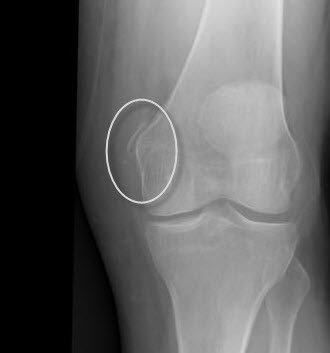
Imaging

The standard anteroposterior x-ray would show a linear soft tissue opacity medial to the femoral condyle. The linear fashion of ossification is often suggestive of calcification of the proximal aspect of the medial collateral ligament. The calcification may also involve the adductor magnus tendon.

Pellegrini Stieda Syndrome Lesion

Computed tomography would also show heterotopic ossification of the femoral attachment of the medial collateral ligament.

MRI would reveal a corticated structure within the proximal MCL, with no evidence of fat content.

MRI is invaluable in defining the anatomy of the structures and confirmation of the site of calcification.

Treatment  of Pellegrini Stieda Syndrome

Nonoperative measures include rest and use of ice massage after activity. Analgesic medications like NSAIDs can be used. Strenuous activity should be avoided.

When conservative treatment is not successful, surgery can be considered. Surgical treatment consists of excision of the bony fragment with careful repair of the medial collateral ligament.


Sports Injuries and Rehabilitation This article has been medically reviewed by Dr. Arun Pal Singh, MBBS, MS (Orthopedics)

About Dr Arun Pal Singh

Dr. Arun Pal Singh is a practicing orthopedic surgeon with over 20 years of clinical experience in orthopedic surgery, specializing in trauma care, fracture management, and spine disorders.

BoneAndSpine.com is dedicated to providing structured, detailed, and clinically grounded orthopedic knowledge for medical students, healthcare professionals, patients and serious learners.
All the content is well researched, written by medical expert and regularly updated.

Read more....

Primary Sidebar

Know Your Author

Dr. Arun Pal Singh is an orthopedic surgeon with over 20 years of experience in trauma and spine care. He founded Bone & Spine to simplify medical knowledge for patients and professionals alike. Read More…

Explore Articles

Anatomy Anatomy Fractures Fractures Diseases Diseases Spine Disorders Spine Disorders Patient Guides Patient Guides Procedures Procedures
featured image of gower sign for segmenatal instability of lumbar spine

Clinical Tests for Lumbar Segmental Instability

Lumbar segmental instability may not always be visible on standard …

mesurement of scoliosis for braces

Braces for Scoliosis- Types, Uses and Results

Braces for scoliosis are recommended to prevent the scoliotic curve …

discogenic back pain

Discogenic Back Pain Causes, Diagnosis and Treatment

Discogenic back pain is a common cause of axial low back pain [the …

Elbow arthrodesis using internal fixation

Elbow Arthrodesis- Indications, Methods and Complications

Elbow arthrodesis refers to the surgical fusion of the elbow joint. It …

skeletal traction in upper tibial pin

Skeletal Traction – Indication, Uses and Complications

Skeletal traction is a type of traction where the force is applied …

Popular articles

Hendrich Fall Risk Score

Currently used scoring system is …

osteopetrosis of femur with fracture, Note the obliterated medullary cavity

Osteopetrosis or Marble Bone Disease

Osteopetrosis is also called stone bone …

Brachial Plexus Injury

Brachial Plexus Injury – Causes, Presentation and Treatment

Brachial plexus injuries can occur in …

lumbar discography in lateral position featured image

Lumbar Discography – Indications, Procedure and Inference

Lumbar discography is a diagnostic …

Bone and Spine

© 2025 BoneAndSpine.com · All Rights Reserved
The content provided on BoneAndSpine.com is intended for informational and educational purposes only. It is not a substitute for professional medical advice, diagnosis, or treatment. Read Disclaimer in detail.