Last Updated on November 20, 2019
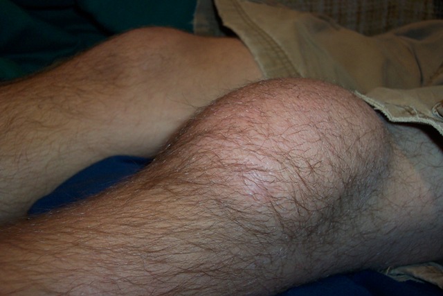
Prepatellar bursitis is inflammation and swelling of the prepatellar bursa which lies between the skin and the patella separating the patella from the patellar tendon and skin. Normally, it contains a minimal amount of fluid and is not appreciable but with inflammation, it could swell substantially.
Prepatellar bursitis is also known as the beat knee, carpet layer’s knee, coal miner’s knee, housemaid’s knee, rug cutter’s knee, or nun’s knee. [All these professions require repeated and prolonged kneeling and the condition was commonly found in these persons]
Noninfective prepatellar bursitis usually affects adults and is more common in females. In children, the cause usually is the infection.
Causes of Prepatellar Bursitis
Usual causation of the prepatellar bursitis is trauma to the anterior aspect of the knee. Trauma could be
- Direct trauma – fall on the knee or direct blow to the knee on anterior aspect.
- Repetitive injury to the knee due to overuse of knee [repeated kneeling]. Commonly found in professions like carpet layering, coal mining, roofing, plumbing, and housemaids.
Infection [in children mostly], crystal deposition diseases [gout or pseudogout] or inflammatory diseases could also lead to prepatellar bursitis.
Presentation of Prepatellar Bursitis
Knee pain with swelling is the most common presenting complaint. The patient might find it difficult to walk and kneel on the affected side. There might be a history of excessive kneeling due to occupational reasons. Sometimes, a history of blunt trauma could be elicited.
On examination, the anterior aspect of knee is swollen [fluid filled swelling] towards the lower pole of patella and redness could be present.
Patella is tender on palpation and crepitation may be elicitable.
Differential Diagnoses
- Cellulitis
- Connective-tissue disorders
- Pes Anserinus Bursitis
- Rheumatoid Arthritis
Lab Studies
Laboratory studies are not usually indicated to diagnose prepatellar bursitis. Fluid aspiration may be sent for ruling out infection or other crystal diseases
Infection is marked by an increase in WBC count of the aspirated fluid, elevated protein, elevated lactate and decreased glucose levels. The fluid may be found positive for bacterial staining. The fluid could be subjected to culture and sensitivity if the findings are suggestive of infection
Crystals in the aspirated fluid could differentiate between different crystal diseases.
- Monosodium urate crystals – gout
- Calcium pyrophosphate – pseudogout
- Cholesterol crystals – rheumatoid bursitis
Imaging
Radiographs are generally not required but may be done to rule out other conditions.
CT scan and MRI may be done in cases where the treatment is not successful.
Ultrasound may rule out intraarticular fluid.
Treatment of Prepatellar Bursitis
Non-septic prepatellar bursitis
- Rest
- Ice
- NSAIDs
- Aspiration of the prepatellar bursa and instillation of corticosteroid [ infection must be excluded]
- Kneepad or cushion
- Physical therapy – Range of motion exercises
- Arthroscopic bursectomy [excision of bursa] in patients who do not respond to nonoperative treatment or have recurrent prepatellar bursitis.
Septic bursitis
RICE therapy along with antibiotics [iv or oral as needed] is usually able to control the symptoms and treat septic prepatellar bursitis
Incision-drainage and/or bursectomy may be required if symptoms of septic bursitis have not improved significantly within 36-48 hours of antibiotic treatment.
Prevention of Prepatellar Bursitis
To prevent the condition, the person must understand the role of recurrent trauma and take protective measures by avoiding frequent kneeling. Protective knee pads can also help.