Sacral fractures are common in pelvic injuries and associated with 30-45% cases. About a quarter of sacral fractures are associated with neurologic injury. Sacral fractures are missed quite often, as often as 75% in patients without a neurological deficit and about half of those with the deficit.
Neurologic injury associated with sacral fractures can range from an incomplete injury of a single nerve root to involvement of the entire cauda equina. The injury can range from a neuropraxia to transection of nerve roots, or even complete transection of the cauda equina.
Sacral fractures either result from high energy trauma in young adults or occur as insufficiency fractures in the elderly as a result of low energy falls.
Sacral fractures are usually associated with pelvic fractures. The sacral fracture associated with lateral compression pelvic fractures is usually stable but sacral fractures associated with vertical shear pelvic fractures are usually unstable. Sacral fractures may involve injury to the lumbosacral junction and result in varying degrees of lumbosacral instability or even lumbosacral dissociation.
L5 nerve root runs on top of the sacral ala whereas S1-S4 nerve roots are transmitted through the sacral foramina. S2-S5 nerve roots perianal sensation and function to control anal sphincter tone and voluntary control. In addition, these function to maintain bulbocavernosus reflex.
Detailed anatomy of sacrum can be read here.
Classification of Sacral Fractures
Denis classification
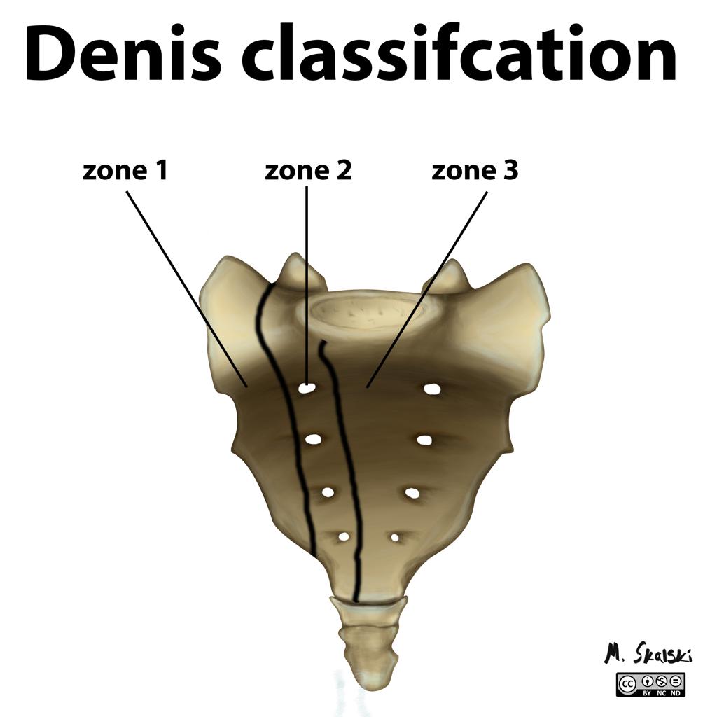
Zone 1
Zone 1 fractures are sacral fractures lateral to foramina. These are most common fractures and associated with the nerve injury in 5%. Usually, the L5 nerve root is affected.
Zone 2
These are the fractures through foramina. These fractures could be stable or unstable and are associated with an increased risk of nonunion and poor functional outcome
Zone 3
These fractures are medial to foramina and involve the spinal canal. These fractures have the highest rate of neurologic deficit [up to 60%] including bowel, bladder, and sexual dysfunction.
Transverse sacral fractures occur in <5% of sacral fractures. They are classified as Denis 3 as they traverse the spinal but the fracture line often traverses all 3 zones.
They have been further classified on morphological pattern H, U, lambda and T-shaped fractures.
Another classification is Roy-Camille/Strange-Vognsen and Lebech Classification of Transverse Sacral Fractures.
Type 1
There is a kyphotic angulation at the fracture.
Type 2
Fractures have both kyphosis and partial anterior translation.
Type 3
Fractures have kyphosis along with a complete translation.
Type 4
Fractures have segmental comminution of the S1 body due to axial compression
Isler Classification
This classification is of fractures involving the lumbosacral junction and is based on the location of the major fracture line on relation to the L5-S1 facet [which impacts the potential for lumbosacral instability.]
Type I
Fractures of the lumbosacral junction lateral to the facet. These generally do not affect lumbosacral stability. Pelvic ring stability may be affected.
Type II
Fractures of the lumbosacral junction that exit through the L5-S1 facet.
Type III
Fractures of the lumbosacral junction that exit medial to the facet. Type III fractures are usually associated with significant instability of the lumbosacral junction. Bilateral type III injuries may represent lumbosacral dissociation.
Presentation of Sacral Fractures
The patient is usually a case of high energy trauma [ motor vehicle accident or fall from height]. Elderly patients may present with sacral fractures after a simple fall.
There is a pain in the posterior aspect of the pelvis. A presence or suspicion of fracture of the pelvis should alert the examiner to look for sacral fractures. All the examination for pelvic fractures must be done. In elderly patients with insufficiency, fracture tenderness may be elicited on the sacrum.
Therefore clinical examination of patients sustaining sacral fractures requires more than the just routine examination of lower extremity sensory and motor function. An additional examination is required to identify injuries to the lower sacral plexus.
This includes
- Rectal exam
- Light touch and pinprick sensation along S2-S5 dermatomes
- Perianal wink
- Bulbocavernosus reflex
- Cremasteric reflexes
Distal pulses should be examined.
Imaging
Xrays are the first line of imaging but about one third are able to show the fractures
Recommended views are Pelvis AP, inlet and outlet views. Inlet view provides the best assessment of the sacral spinal canal and superior view of S1. Outlet view provides a true AP view of the sacrum.
In addition, lateral view of sacrum can provide more information on sacral fractures as well as a lumbosacral junction.
CT is the imaging choice to identify the fracture pattern better. Coronal and sagittal reconstruction enhances understanding.
MRI is recommended for assessment of neural deficit.
Treatment of Sacral Fractures
Nonoperative Treatment of Sacral Fractures
Most sacral fractures can be treated nonoperatively. To treat nonoperatively the fracture should be
- Undisplaced/stable
- Without a significant associated pelvic ring disruption
- Not involving the lumbosacral junction
- Without neurologic injury.
Nonoperative treatment involves limitation of weight bearing with a gradual increase, orthosis.
Operative Treatment
Surgical fixation of sacral fractures is indicated in
- Fractures with displacement>1 cm
- Soft tissue compromise
- Persistent pain after non-operative management
- Displacement of fracture after non-operative management
Decompression is also indicated in fractures with the neural deficit.
Sacral Fracture Fixation
Displaced or unstable sacral fractures are best treated by closed or open reduction and internal fixation. Following types of fixations are used. Neural decompression when needed, may be achieved indirectly by fracture reduction, or a laminectomy or foraminotomy.
- Percutaneous Iliosacral Screws – most commonly used
- Posterior Sacral Tension Band Fixation – Vertically unstable comminuted sacral fractures or patients with a dysmorphic sacrum
- Lumbopelvic Fixation – In sacral fractures with associated lumbosacral instability [Pedicle screws placed in L5 and or L4 that are connected to fixation placed into the ilium from posterior to anterior.]
- Triangular Osteosynthesis – Placement of pedicle screws in the lower lumbar and the posterior ilium in conjunction with iliosacral screws.
Complications of Sacral Fractures
- Venous thromboembolism due to immobility
- Iatrogenic nerve injury
- Malreduction
- 21Shares
21
I had a s1 s2 sacral ala fracture from a fall. I am 48 years old and pretty healthy. Is it normal for the follow up CT scan at 6 1/2 weeks to show no evidence of bony bridging yet? How long does that normally take for the fracture line to fill in?
Joanne,
Should start showing signs by now. Some people heal slowly. Wait for another month or so before getting another image.
All the best.
SIR
This is the CT Scan report of my son who had an accident 4 days back. He has full bladder control , but hasnt opened bowels yet. Will he have any problem ?
He will be discharged in 2 days. Can you please suggest how to nurse him
Undisplaced fracture in the left transverse process of L5 vertebra.
Comminuted undisplaced fracture in the left lateral part of the sacrum involving all the sacral vertebrae. Comminuted undisplaced fracture of the right lateral part of the sacrum involving the S1 vertebra.
Comminuted undisplaced fractures in the right pubic bone, right superior pubic ramus and anterior pillar of the right acetabulum.
Comminuted undisplaced fracture of the anterior pillar of the left acetabulum and the left inferior pubic ramus.
Normal alignment of the lumbar vertebrae are maintained
The intervertebral discs appear normal. No obvious disc bulge or herniation is seen.
Rest of the vertebral bodies, pedicles, transverse processes, laminae and spinous process appear normal. No obvious sclerotic or lytic lesion is seen.
The facet joints and neural foramina are within normal limits.
The thickness of ligamentum flavum is normal at all the levels.
Both sacroiliac joints appear normal.
The paraspinal soft tissue does not show any abnormality.
Gayathri,
If bladder has returned, bowel would follow too. As for as nursing is concerned, you would need to learn from your treating facility.
Take care.
Hi doctor,
I had a fall 13 days ago and MRI result finding is as follow. I have difficulty sitting, laying down on my butt, even standing at times. Bowel movements are also difficult like I have been constipated.
MRI
The comminuted mildly displaced fracture of the S4 segment is seen, with discontinuity of the anterior cortex, extensive adjacent T1 hypointense and STIR hyperintense marrow edema and mild presacral swelling. there is more extension of the edema and probable fracture to the right sacral wing. Compression of the sacral spinal canal is noted.
Kelly,
How could I be of help. I ask this because you did not ask anything. How are you doing?
Take care.
Oh, what could possibly be the treatment and what could happen to me? Currently pending CT scan. Possible surgery? How long does it take to heal? What will be restrictions? What can I do to heal faster? Pls advise. Thank you!
Hi Doctor,
I had a mildly displaced lateral fracture between S3 – S4 after a fall.
I’ve experienced some nerve compression resulting in bowel control issues.
It’s been 12 weeks and I’m still experiencing discomfort and pain.
Is this to be expected this long after the accident?
I’m due to visit the hospital in 2 weeks but am unsure if the medical staff plan to CT or MRI scan me. Should I ask for this to happen?
Many thanks,
Paul
Kelly,
Whether you need surgery or not would be determined by the clinician who sees you. There would be restrictions on movements and sitting as determined for your injury.
Eat well and do not take stress for better healing.
Paul Styles,
You should discuss it in detail if there is a nerve compression being caused by your injury [ If there is a loss of control of bowel, it could be cause].
An MRI would help to know the status better.
Take care.
Hi, I had a S1, S2 sacral ala fracture from a fall. It’s now been 4 months since the accident but I am still experiencing mild pain in the sacrum and L5 (which showed degenerative changes). Will this be a chronic pain that I have for the rest of my life or is it more likely that patients have a full recovery?
Thank you
Kristina,
Most of the people become better. Chronic pains if likely come after pretty long time. Please ask your treating doc abotu the prognosis of your injury.
Take care.
Doctor
I have been fused from L2-L5 for 4 years. Also 5 cervical fusions. I developed sever burning pain in my hips running thru my croch. I now have osteopenia. Finally, had MRI if sacrum. I have 4 insufficient fractures involving bilateral inferior sacrum, coccyx, bilateral posterior ilium and bilateralacetabular roof. What zones are these? What does all this mean?
Insufficent fractures are fractures resulting from less than normal force required to break the particular bone. Bones weakened by osteopenia could be a cause. Other may be deficiency of vit D etc . You need to speak to your physician if the causation is to be established.
You would understand the regions mentioned here in your report by looking at a diagram of pelvis. Just to get an idea. Ilium is hip bone, coccyx is last part of spine, acetabular roof is the area of hip bone just above the hip joint.
I hope that helps.
hi , 16 weeks ago i fractured my sacrum undisplaced involving s3-s5 segments im still really sore and burning pain but my mobility is heaps better.
im 43 and fell down a timer staircase
Would an SI belt work to help with pain for a pt with a stable sacral alar fx?
Jerry,
It should help by providing immobilization in flexion and extension plane and providing structural support.
Thanks for the query.
Two weeks post postpartum injury, six weeks immobile with horrific pain. Injury remained undiagnosed until 3 years later when rolling over in bed resulted in similar immobility and pain.
Diagnosis of left inferior sacral shear with torqued pelvis’was made upon physical manipulations, verified by CT.
Longer leg on the left, due to share.. Subsequent L4 and L5 disc herniations intermittent treated by traction in hospital and out patient.
Injury is now 33 years old, in a mostly active 65 year old woman, with episodes of pain and carrying degrees of immobility occurring with increasing frequency.
* Would lifts in the short leg side’s shoes help with ability to walk further without pain?
* What is an expected prognosis for future mobility issues?
For now reliance upon a wheelchair is sometimes necessary, sometimes merely a came, and the pain and mobility issues, tho increasing in frequency remain intermittent.
Thank you in advance of your consideration
Moon,
if there is a significant shortening, the shoe raise does help. But the pain is often not due to shortening per se but the changes that follow. The amount of relief would depend if there are correctable changes or not.
I cannot comment on prognosis given the limited data and interface limitation. You would need to ask your treating doctor.
Take care.
My mother is 88 yrs old.was just diagnosed with a visual left sacral insufficent fracture, She does have osteoporosis. What can we do for her? Will this require surgery or lots of physical therapy. She is also a recovering bladder cancer survivor and has a urostomy. I’m concerned about surgery because of her age and possibly re-fracturing because of her lack of bone density.
Martha,
While the specific answer would be given by the treating doctor, insufficient sacral fractures should generally heal with rest and medication.
I hope that helps.