Last Updated on October 29, 2023
Synovectomy knee is a surgery where synovium of the knee joint is removed surgically. Synovectomy of knee classically was done with open incision but with increasing success of arthroscopic synovectomy, arthroscopic synovectomy of knee offers an option where there is minimal soft tissue trauma. The goal in synovectomy is total synovectomy which means complete removal of synovium but for all practical purposes, most of the synovectomies are subtotal.
Indications for Synovectomy of Knee
- Aggressive rheumatoid arthritis,
- Generalized or localized pigmented villonodular synovitis,
- Synovial Chondromatosis
- Infectious synovitis [as in tuberculosis]
- Secondary arthrosynovitis
- Post-traumatic
- Degenerative
Surgical Procedure of Open Synovectomy Knee
For reference to structures, Anatomy of Knee Joint may be read.
Steps of Open Synovectomy Knee
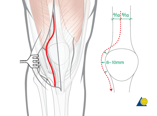
- The incisions
- Midline anterior or
- Medial/lateral parapatellar incision
- Some surgeons prefer two parapatellar incisions.
- Expose from the suprapatellar pouch proximally to the insertion of ligamentum patellae distally
- Skin, superficial fascia, deep fascia are cut
- Quadriceps expansion is cut about half to one cm away from the patella.
- By sharp dissection, a plane of cleavage is developed between the quadriceps expansion and suprapatellar synovial pouch.
- Excise the thickened synovial membrane starting from its proximal borders the suprapatellar pouch, articular margins of femoral and tibial condyles.
- Flex the knee joint to 90 degrees and inspect
- Remove any loose bodies
- Remove as much as an unhealthy synovial membrane that can be removed from the cruciate ligaments and intercondylar fossa.
- Rotating tibia to either side may maximize the exposure of the membrane
- Above maneuver also helps inspection of menisci.
- Any obstructing intra-articular lesion or extra-articular adhesions should be cut’. When required, capsulectomy or capsulotomy is performed, to obtain the desired flexion on the operation table. The goal is to achieve a minimum of 90 degrees of flexion at the knee.
- The wound is closed over suction drainage
In cases of infection, such tuberculosis the disease might have spread beyond the extent of the synovial membrane.
In such cases, debridement of the joint is also undertaken. This involves removal of pannus, curettage of destroyed bone.
If the cavities are large, these may be filled up with cancellous bone grafts from the nearby healthy bone. All the destroyed tissue like menisci, loose and destroyed articular cartilage, destroyed capsule and ligaments may be carefully removed.
If the joint is extensively destroyed (>50%), arthrodesis should be done.
Postoperative Care for Synovectomy Knee
The limb is kept elevated on a pillow with the knee joint kept in about 5 to 10 degrees of flexion with the help of a rolled towel or a small pillow behind the knee.
Exercises of the ankle and static quadriceps exercises are started the same day.
Knee bending exercises are started after 24-48 hours after removal of suction drain. The patient may be put on continues physiotherapy.
Quadriceps strengthening is continued and weight bearing is gradually added.
Arthroscopic synovectomy knee
Arthroscopic synovectomy knee has many advantages over open synovectomy
- Minimally invasive surgery – As compared to long incisions and open dissection, only small incisions for portals are made
- Short hospital stay
- Decreased risk of postoperative joint stiffness,
- Similar results to open synovectomy
Five arthroscopic portals are used in an arthroscopic synovectomy. The number of ports and exact location may vary among surgeons.
- Lateral patellofemoral axillary portal
- High parapatellar anteromedial portal
- Posterolateral portal
- Posteromedial portal
- Superomedial portal.
Using the above portals the synovectomy can be done in the following sequence
- Intercondylar notch to make a gateway for posterior compartments
- Posterolateral compartment
- Posteromedial compartment
- Medial compartment
- Lateral compartment
- Suprapatellar compartment
- Retropatellar compartment.
Some surgeons though prefer to do arthroscopic synovectomy of the knee as performed from the anterior to posterior compartments following the location of anatomical structures.
Clear arthroscopic visualization is crucial for manipulating arthroscopic instruments effectively.
Postoperatively, a bulky dressing is applied.
References
- D.J. Ogilvie-Harris, L. Weisleder.Arthroscopic synovectomy of the knee: Is it helpful? Arthroscopy, 11 (1995), pp. 91–95
- K. Schmidt, R.K. Miehlke, R. Rupprecht.Open or arthroscopic synovectomy of the knee in rheumatoid arthritis.Aktuelle Rheumatol, 20 (1995), pp. 212–220
- Kim SJ, Jung KA, Kwun JD, Kim JM. Arthroscopic Synovectomy of the Knee Joint in Rheumatoid Arthritis: Surgical Steps for Complete Synovectomy . Arthroscopy Volume 22, Issue 4, April 2006, Pages 461.e1–461.e4. doi:10.1016/j.arthro.2005.06.025