Last Updated on November 19, 2019
Tuberculous of the hip joint is common with a frequency of involvement next only to spinal tuberculosis. Hip tuberculosis accounts for 15% percent of all cases of osteo-articular tuberculosis and 1-3 % of all tuberculosis cases.
Anatomy of the Hip Joint
The hip joint is a ball and socket synovial joint. Fibrocartilaginous labrum attached to the acetabulum makes the socket deeper, however, a considerable part of the articular surface of the spherical femoral head remains uncovered.
The opening of the acetabulum faces laterally, downwards (about 30 degrees), and forwards (about 30 degrees). Consequently, the femoral neck points medially, upwards and anteriorly.
The forward inclination or the angle of anteversion in an adult is 10 to 30 degrees. The neck-shaft angle is 125 degrees.
A neck-shaft angle of less than 125 degrees is referred to as coxa vara and the angle more than 125 degrees (i.e. more vertical neck) is called coxa valga.
Coxa vara causes limitation of abduction and internal rotation due to mechanical reasons.
During standing and walking the femoral neck acts as a cantilever
The femoral head receives its arterial blood supply from 3 sources
- Intraosseous vessels running up the neck
- Vessels in the retinacula reflected from the joint capsule onto the neck.
- Vessels in the ligamentum teres
The capsule of the hip joint is attached circumferentially around the labrum acetabulum and transverse ligament
The fibrous capsule is strengthened by 3 ligaments
- lliofemoral ligament(Y-shaped ligament of Bigelow)
- Pubofemoral ligament
- Ischiofemoral ligament
The synovial membrane lines the inner surface of the whole of the capsule and is reflected proximally along the reflection of the capsule investing the retinacular fibers up to the articular margin of the femoral head.
Anteriorly the iliac bursa lies over the capsule and extends upwards into the iliac fossa beneath the iliacus muscle. The psoas major tendon and iliacus muscle separate the capsule from the femoral vessels and femoral nerve.
Superiorly there is a cellular space between the capsule and the overhanging gluteus minimus and piriformis muscles.
Inferiorly the obturator externus muscle spirals back around the femoral neck.
Posteriorly the obturator internus and gemelli separate the sciatic nerve from the capsule. Medially the thin acetabular fossa forms part of the lateral wall of the pelvis.
Read Detailed Anatomy of Hip Joint
Pathophysiology of Tuberculosis of Hip
Tuberculosis of hip, like other osteoarticular tuberculosis, is secondary to primary pathology in lungs, lymph nodes or any of the viscera. Through the hematogenous route, the bacteria reach either to synovium or bone.
The disease process starts in
- Acetabular roof [most common]
- Epiphysis of the head of the femur
- Metaphyseal region
- Greater trochanter
- Synovial membrane [rarely]
Isolated involvement of greater trochanter and trochanteric bursa without the involvement of hip joint is known.
In other cases of origin of the disease, the hip joint gets involved quickly to become an osteoarticular disease.
The involvement of the joint leads to the destruction of articular surfaces of femoral head and acetabulum. Because, the disease starts slowly and symptoms are mild initially, the patient often presents late and by the time diagnosis is reached at, the joint is destroyed to quite an extent.
An abscess [cold abscess] is formed within the joint, may perforate the inferior weaker part of the capsule to present as cold abscess anywhere around the hip.
It may also perforate the acetabular floor and become an intrapelvic abscess.
Symptoms and Signs of Tuberculosis of Hip
Hip tuberculosis most commonly present in first three decades though no age is immune. The commonest decade of presentation is the third decade. The patient usually presents with pain and limping and deformity around the hip.
Pain may be referred to medial aspect of the knee. The severity of the pain is maximum at the end of the day.
The limp is the earliest and commonest symptom. The patient, in an attempt to put as little weight for as little time on the affected limb, shortens the stance phase and walks with an antalgic gait.
Turning in the bed may be difficult and the patient might support the limb with contralateral normal limb when attempting to lift the affected limb.
On examination, the hip joint would be tender. There would be muscle spasm in lower abdominals and adductors of the hip and disproportionate wasting of muscles of the affected limb.
Natural History of the Hip Joint Tuberculosis
In its natural history, tuberculosis of the hip passes through different stages, depending upon the extent of involvement and amount of destruction.
Stage 1 – Tubercular Synovitis
Synovitis stage occurs when the lesion is in the early stage. Typically, the lesion is juxta-articular osseous lesion which causes irritation of the hip.
The joint is held in the position flexion, external rotation and abduction to increase the capacity of the joint.
This position leads to apparent lengthening but there is no change in the true length of the limb.
The joint is painful only in extremes of movement.
X-rays may be entirely normal or show soft tissue swelling. Rarefaction of the hip bones may be present.
Stage 2 – Early Arthritis
This is an advanced stage, there is damage to bone and cartilage of the joint. There occur adductor and flexor spasm leading to flexion, adduction and internal rotation deformity of the joint.
This presents as an apparent shortening of the limb. There might be some amount of true shortening but apparent shortening appears more.
Muscle wasting becomes appreciable and there is a restriction of movements of the hip in all directions.
There is osteoporosis of the involved bones. This occurs due to the increased circulation of the bone following inflammation. A slight decrease in the joint space may be visible due to cartilage destruction.
Stage 3 – Advanced Arthritis
There is the destruction of cartilage and bone and, the signs present in stage 2 are exaggerated. On x-ray, there is a further narrowing of the joint space.
Stage 4 – Advanced Arthritis with subluxation or Dislocation
There is a destruction of the acetabulum, femoral head, capsule, and ligaments. This may lead to the displacement of the femur upwards and dorsally in the widened acetabulum.
Frank dislocation of the head of the femur may also occur.
in the wandering or migrating acetabulum leaving its lower part empty and Shenton’s arc broken.
Rarely the destruction of capsule and acetabulum may be so severe as to lead to frank pathological posterior dislocation of the femoral head.
The acute variety of tuberculous infection can rarely be encountered in children. Sometimes the hip may show protrusion through the acetabulum.
In some cases, the femoral head and neck are grossly destroyed, collapsed and small in size contained in an enlarged acetabulum-mortar and pestle appearance.
All the movements of the hip are grossly restricted. Some cases where acetabulum is widened [wandering acetabulum, protrusio acetabuli or mortar and pestle picture may retain a fairly good range of movements for a long time.
Classically described deformity is flexion, adduction and internal rotation. But some cases may have with abduction and external rotation. This occurs either because of keeping the limb in that posture for rest or their destruction of iliofemoral Y ligament.
Diagnosis of Tuberculosis of Hip
In the endemic regions for tuberculosis, a clinical diagnosis supported by radiographs is considered adequate for starting the treatment but where the disease is not common, additional investigations like ultrasound, MRI or biopsy may be needed.
Xray changes are often late and confirmation of diagnosis by the demonstration of tubercular bacilli or histopathological examination is recommended.
In advance stages of arthritis, radiological changes are typical and tissue diagnosis may not be required.
Imaging Studies
Xrays
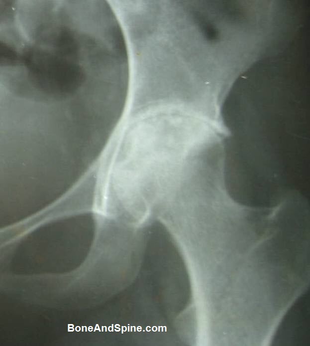
Initial X-rays may be normal. Decreased joint space and osteoporosis of the bones is found in advanced stages. The lytic lesion may or may not be evident on x-ray.
Sometimes two lytic lesions are visible on either side of the joint and called kissing lesions.
In the advanced arthritic stage, there may be a break in the Shenton line [visible on xray], and extreme reduction of joint space.
In long-standing cases, secondary arthritic changes such as osteophytes may be seen.
Classification of the Radiological Appearance
Shanmugasundaram suggested a new radiological classification for the tuberculous disease of the hip applicable for the lesions in children(C) and adults(A).
Following radiological types are suggested by him
- Normal appearance (C)
- Traveling acetabulum (C, A)
- Dislocated hip (C)
- Perthes type (C)
- Protrusion acetabuli (C, A)
- Atrophic type (A)
- Mortar and pestle” type (C, A).
There is a relationship between various radiological types and the functional outcome.
- Good results were obtained in
- 92 percent of ”normal type”
- 80 percent of Perthes’ type
- 50 percent of dislocating type
- 29 percent of traveling -acetabulum and Mortar pestle type.
The poor outcome could be predicted if the joint space was reduced to 3 mm or less.
MRI
MRI shows the predestructive lesion like edema and inflammation and is a sensitive test to detect soft tissue abnormalities but is not specific.
MRI in early stages may show synovial effusion and varying degree of bone edema, minimal areas of bone destruction. MRI is very useful in the early stages of the disease where the x-rays are normal. These may show synovitis, destruction of cartilage and the presence of fluid in the joint.
Lab Studies
Routine lab studies are not specific for tuberculosis but may point towards the presence of inflammation.
CBC, ESR, and CRP are routine studies. There may be an increase in the number of lymphocytes. ESR and CRP are raised but there are also cases where these indices are not changed.
Mantoux Test may be done but does not have any value in the endemic region
Tissue Diagnosis
In cases where a diagnosis cannot be made, tissue can be obtained from the hip joint, either by open biopsy or arthroscopic biopsy. The synovial biopsy may give a conclusive diagnosis where clinical diagnosis is equivocal.
The obtained tissue is subjected to the following tests
- Histopathological examination
- Acid-fast Bacilli [AFB staining]
- Polymerase chain reaction for tuberculosis
Differential Diagnoses of Tuberculosis of Hip
The stage of the disease might guide the differentials as well.
- Traumatic synovitis,
- Rheumatic/rheumatoid disease
- Transient synovitis of the hip
- Perthes’ disease
- Juxta-articular disease-causing irritation of the joint
- Spasm of the iliopsoas muscle due to
- Abscess
- Lymph node
- Slipped capital femoral epiphysis.
- Osteomyelitis of the upper end of the femur
- Hip injury
- Acute bacterial arthritis
- Osteoid osteoma of the neck of the femur
- Villonodular synovitis
- Avascular necrosis of the head of the femur
- Giant cell variants of upper femur
Treatment of Tuberculosis of Hip
The treatment of the tuberculosis of hip joint aims at
- Control of active infection
- Correction of deformities
- Restoration of function
Antitubercular Chemotherapy
Standard chemotherapy as accepted universally with four antitubercular drugs, namely isonizid, rifampicin, pyrazinamide, and ethambutol to start with in all the stages of presentation is recommended.
Traction
Along with chemotherapy traction is recommended to all patients. Skeletal traction is preferable to skin traction. In the presence of abduction deformity, bilateral traction is mandatory.
Traction serves the following advantages
- Relieves the muscle spasm
- Prevents or corrects deformity and subluxation
- Maintains the joint space
- Minimizes the chances of development of migration of acetabulum
- Permits close observation of the hip region
- Keeps the joint surfaces apart early hip mobilization exercises can be started.
Classically, weight-bearing was allowed after 4-6 months of treatment but in modern practice, earlier weight-bearing, whenever the patient can tolerate the pain is recommended.
Surgery
Synovectomy, Joint Debridement, Arthrolysis
These measures are generally taken in early and late arthritis. The aim is to decrease the tissue load and break the adhesions for a better outcome of the treatment.
Excision Arthroplasty
In the populations where people are used to floor level activities and positions such as squatting, sitting cross-legged and kneeling excision arthroplasty are one of the surgical treatments.
Also called Girdlestone’s excision arthroplasty, the surgery can be safely carried out in healed or active disease after the completion of the growth potential of bones of the hip joint.
The procedure involves the removal of upper end of the femur, followed by heavy traction so that fibrous tissue is formed and postoperative in bed mobilization to get a mobile hip. Some amount of shortening occurs
Girdlestone procedure provides a mobile, painless hip. About 90% of patients are able to squat and kneel and able to sit cross-legged and walk with help of a stick.
Arthrodesis
Arthrodesis of the hip, though relieves the pain, and corrects the deformity but sacrifices the motion. Arthrodesis as a procedure is done less frequently now due to increasing acceptance of total hip arthroplasty.
Corrective Osteotomy
Patients presenting with sound ankylosis in bad position require upper femoral corrective osteotomy.
Hip replacement
Total hip arthroplasty in the hip with active tuberculous infection is a controversial issue due to the potential risk of reactivation of TB. But acceptability of early total hip arthroplasty is gradually increasing globally with reports that metal can be safely used in tuberculous lesions.
Stagewise Treatment Approach
Synovitis
- Analgesics
- Antitubercular chemotherapy
- Traction, rest and mobilization
- Surgical interventions usually are not required
Early arthritis
- Treatment similar to stage
- Synovectomy and joint debridement – to reduce the disease tissue load
- The prognosis, in general, is good.
Advanced arthritis
- Treatment in stage 2
- Arthrolysis of joint with joint debridement.
Advanced arthritis with subluxation/dislocation
Joint destroyed, deformities and shortening present
- Chemotherapy, analgesics, traction
- Arthrodesis/Excisional arthroplasty/ total hip arthroplasty
Complications of Tuberculosis of Hip
- Premature closure of knee growth plates in prolonged immobilization in plaster [>12 months]. This could lead to marked shortening of the limb. Such kind of immobilization is rarely done now.
- Coxa magna and valgus hip due to increased circulation of the region
- Coxa vara – Rarely occurs following a destructive lesion in the femoral head and neck
- Rarely simultaneous damage to the trochanteric physis and femoral head physis would result in generalized hypoplasia of upper end of the femur
- Active disease during growing age may interfere with the blood supply of the epiphysis of the femoral head giving rise to radiological picture resembling Perthes’ disease
Prognosis of Hip Tuberculosis
- Early disease (synovitis or early arthritis) may heal leaving a normal or nearly normal hip joint.
- Advanced arthritis generally results in fibrous ankylosis.
Without and treatment – flexion adduction deformities develop. Require some kind of surgical procedure
Management of Tuberculosis of Hip in Children
In children, late secondary osteoarthritis is always a concern. Moreover, the deformities in one hip would affect the other hip as well as spine and knee.
Therefore, early detection and treatment is even more important in this age group.
In children, once the tuberculosis is suspected clinically, radiological examination of both hips and lungs should be done.
Some authors recommend ultrasound assessment of hip to gauge if a drainage is required.
Synovitis and early arthritis are treated by nonoperative measures. Arthrotomy and debridement may provide quicker response.
In very young children hip spica can be applied for 4-6 weeks instead of traction.
Children have great potential for regeneration and remodeling. Therefore excision arthroplasty or arthrodesis is not indicated before skeletal maturity.
In case of wandering acetabulum, an early osteotomy can be done.