Last Updated on September 5, 2020
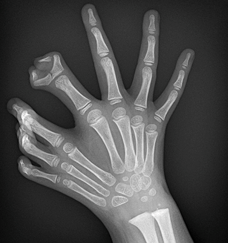
Ulnar dimelia is also called mirror hand syndrome. Ulnar dimelia is a congenital deformity where the radial ray, [the radius, scaphoid, trapezium, first metacarpal, and the phalanges of the thumb] are absent and there is a duplication of ulnar ray [una bone, carpal other than those in radial ray, metacarpal and phalanges of 2nd-5th finger]. There appear seven or eight fingers in the hand.
The abnormality may be associated with duplication of the feet. Ulnar dimelia is usually not hereditary and quite a rare condition.
The skeletal deformity may be accompanied by neural and arterial abnormalities such as
- Duplication of the ulnar nerve
- Duplication of the ulnar artery
- Abnormal arterial arches
- Shortening of the radial nerve,
- Absence of the radial artery
Ulnar dimelia is also known by name. of mirror brush

Pathophysiology
The ulnar dimelia is considered a type III congenital deformity of hand which means it’s a deformity of duplication.
During fetal development of upper limb, is the rise of limb buds, also called Wolff crest which appear on the ventrolateral side of the embryo.
The bud’s distinct upper or cranial margin is called the preaxial border, and the lower or caudal margin is called the postaxial border.
Thus, with the adult upper limb in the anatomical position, the preaxial border runs from the tip of the shoulder to the thumb. The postaxial border runs from the base of the axilla (armpit) to the little finger.
The limb buds are formed by mesenchyme [forms connective tissue] covered with a layer of ectoderm [forms skin and appendages].
The apical ectodermal ridge (AER) is a structure at the distal end of each limb bud which is responsible for induction and modulation of the limb growth.
The mesoderm of the limb buds is divided into the zone of polarizing activity (ZPA) proximally and a progress zone (PZ) distally.
The ZPA mesenchyme inducted cartilaginous and muscular tissue.
The disturbance of differentiation may lead to post-axial duplication and ulnar dimelia.
HOX genes mutations at chromosome 7, 17, 12 have been suggested to be involved. Translocation breakpoint at 14q has also been found.
There are two types of ulnar dimelia
- Type I
- Duplication of ulna
- 5 proximal carpal bones – 2 triquetral, 2 pisiform and 1 lunate
- 5 distal carpal bones- 2 hamate, 2 capitate, and 1 trapezoid
- One index finger in the middle and three other fingers [middle, ring, and little] on both sides of index
- Type II – All others structures are the same as the type I except for the following
- 2 lunates
- 2 trapezoids
- 2 index fingers
Classification of Ulnar Dimelia [Al-Qattan and Al-Thunayan]
There are five types of ulnar dimelia as per this classification
- Type 1
- two ulnae with absent radius
- Type 2
- Two ulnae and a radius
- Type 3
- Mirror hand polydactyly with one radius and one ulna
- Type 4
- a mirror hand with fibular dimelia
- Also called Laurin Sandrow syndrome
- Type 5
- Multiple hands-on the end of the forearm
Clinical Presentation
Usually, only one side is involved. There would be 7 or 8 fingers in the involved hand depending on the type of ulnar dimelia.
On examination, the hand is palmar-flexed at the wrist and radially deviated. The wrist and elbow are broader than normal.
There is a restriction of the motion of the forearm rotation [supination and pronation]. The flexion-extension motion of the elbow is also restricted.
The fingers are held in flexion because of absence or hypoplasia of the extensor digitorum longus muscles.
The intrinsic muscles of the hand are weak. Ulnar fingers tend to be more normal and functional than the radial digits.
Syndactyly of some of the digits may be present.
Metacarpals diverge, and there is a cleft in the palm.
Imaging
Anteroposterior and lateral views are routine x-rays that are sufficient to arrive at a diagnosis of ulnar dimelia. Any associated anomaly is investigated accordingly. The following are the typical x-ray findings.
- Presence of two ulna
- Humerus articulates with two ulnae
- Absent of radial ray i.e.
- Radius
- Thumb
- Duplication of
- Ulna
- Ulnar halves of the carpals and metacarpals
- Presence of eight triphalangeal digits
Treatment
The objective of treatment is to improve function and provide a cosmetically more attractive hand. The management involves a series of surgeries.
The surgical correction is delayed till the age of one year and till that time orthosis may be used if required.
The disease requires surgical intervention for the creation of thumb (Pollicization).
The most normal preaxial digit is chosen for pollicization.
The intervening supernumerary one or two digits are sacrificed and the excess skin is utilized to create a thumb web.
The remaining divergent metacarpals may be osteotomized to close the palmar cleft. A free tendon graft is used to hold the metacarpals together.
References
- Gropper PT. Ulnar dimelia. J Hand Surg. 1983;8A:487–491.
- Yang SS, Jackson L, Green DW. A rare variant of mirror hand: a case report. J Hand Surg. 1996;21A:1048–1051.
- Apiou F, Flagiello D, Cillo C. Fine mapping of human HOX gene clusters. Cytogenet Cell Genet. 1996;73:114–115