Last Updated on October 29, 2023
Vertebral canal or spinal canal is the long tubular space in the vertebral column formed by contiguous placement of vertebral foramen through which the spinal cord passes along with other contents
Vertebral foramina are stacked on one another to form a long canal.
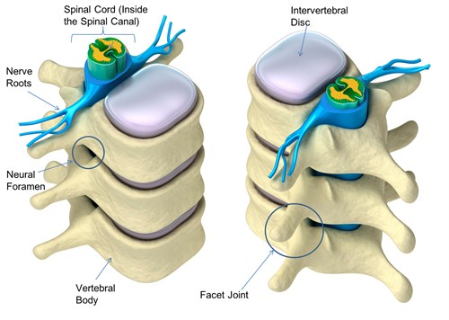
Image credit: NEBH
In the intervertebral spaces, the canal is protected by the ligamentum flavum posteriorly and the posterior longitudinal ligament anteriorly.
Vertebral Foramen and Vertebral Canal
Vertebral foramen refers to empty space enclosed by the ring formed by the neural arch and posterior aspect of the vertebral body.
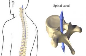
Image Credit: Alberta
When individual foramen are lined over one another, they form a long tubular canal which extends from cervical to sacral region. This is called vertebral canal. [Coccygeal vertebrae are fused and do not contribute]. The dimensions of the vertebral canal vary in different regions of spine depending on the size of vertebral foramen.
Intervertebral Foramen
Vertebral foramen should not be confused with intervertebral foramen. Former is within a single vertebra whereas other is created when two vertebrae are stacked together. Vertebral foramen is a single large hole in vertebra but intervertebral foramen are created on either side between two vertebrae.
Intervertebral foramen allows for passage of nerve roots out from the spinal canal along with spinal artery of the segmental artery, communicating veins, sinuvertebral nerves and, and transforaminal ligaments.
Specifically, the intervertebral foramen is bound by the superior notch of the adjacent vertebra, the inferior notch of the vertebra, the vertebral body, and facet joints on the transverse process of the vertebra.
Contents of the Vertebral Canal
The tubular vertebral canal contains the spinal cord, its meninges, spinal nerve roots, and blood vessels supplying the cord, meninges, vertebrae, joints, muscles, and ligaments.
The canal is enclosed within its column and formed by the juxtaposition of the vertebral foramen, lined up with one another in series. The vertebral bodies and discs make up the anterior wall along with the posterior longitudinal ligament, whereas the laminae and ligamentum flavum border the canal posteriorly. Laterally, spinal nerves and vessels travel through the intervertebral foramen.
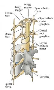
Image Credit: BiologyforISC
Meninges and related spaces
There are three meninges or the layers of membranes which cover the brain, spinal cord, and nerve roots.
Meninges protect spinal cord and roots.
The outermost layer is the duramater which is a resilient layer. The middle layer is arachnoid mater and the innermost layer is piamater. The inner two layers are together are called leptomeninges.
The spinal cord, roots, and nerve rootlets are closely invested by the pia whereas the dura and arachnoid together form a loose sheath called (termed dural/thecal sac) around these structures, separated from the canal walls by the epidural space.
Spinal Dura Mater
The dura is made of tough longitudinal collagen fibers interwoven with circular elastic fibers. The external surface is rough and blends with loose connective tissue in the epidural space. The internal surface, facing into the subdural space, is smooth and covered by a layer of mesothelium.
Inferiorly, the dural sac ends at the sacral canal, usually at S2-S3 or sometimes S1
The dura continues caudally as a fibrous thread named the filum terminale externum or coccygeal ligament, which blends with the PLL over the coccyx.
From dural sac, sleeves are projected into the intervertebral foramen, where the dura blends with the epineurium of the spinal nerves.
Connective tissue slips in the foramen anchor the dural sleeves to protect spinal nerve roots from being stretched during L-spine movements.
Dura is also attached in places to the posterior longitudinal ligament.
Epidural Space
The epidural or extradural space [also termed as peridural space] is a space outside the duramater. The nerve roots transverse the space as they extend into the intervertebral foramen.
This space is occupied by loose connective tissue and fat. It provides padding around the dural sac and spinal cord and acting as a form to hold the thin internal vertebral plexus of veins open. The vertebral venous plexus is embedded in the epidural loose connective tissue, sometimes transmitting large amounts of blood.
Epidural space ends at the sacral hiatus, sealed by the posterior sacrococcygeal ligaments.
Leptomeninges
The pia and arachnoid are composed of loose connective tissue and separated from one another by the subarachnoid space. They are quite delicate membranes.
They are covered by a layer of mesothelium bathed by cerebrospinal fluid.
The arachnoid mater lines the entire dural sac and extends into the dural sleeves. Trabeculae formed by this membrane in subarachnoid space facilitate CSF mixing.
Inferiorly, arachnoid lines the dural sac within the sacral canal and ends on termination of the sac at the S2 level.
The pia mater provides support for the vasculature and nerves in the subarachnoid space. It adheres intimately to the spinal cord.
Pia mater forms a separate sheath for each root and blends with epineurium.
It continues as filum terminale internum caudally and finally becomes enclosed within the filum terminale externum and continues to the coccyx.
Subarachnoid Space
Subarachnoid space is quite spacious in the lumbar area. It is called the lumbar cistern below L2. Subarachnoid space contains cerebrospinal fluid. Spinal block is given by injecting drugs into this space. Lumbar puncture is also done by injecting the needle into this space.
Spinal cord
Brain and spinal cord are the two main components of the central nervous system. The spinal cord functions as a reflex center and conduction pathway between the brain and the body.
Spinal cord starts below brain stem and terminates as the conus medullaris within the lumbar spinal canal at the lower margin of the L2 vertebra.
Different levels of the spinal cord are responsible for supplying different areas of body and organs.
Vertebral canal and spinal cord have different growths and different lengths. Therefore vertebral and spinal levels are different.
Spinal Nerves and Roots
Dorsal roots are somatosensory which arise from the posterolateral aspect of the spinal cord and a ventral or anterior are the motor which arises from the anterolateral aspect of the cord. They join in the spinal canal to form the spinal nerve root which then exits at their respective neural foramina as spinal nerves.