Last Updated on October 11, 2023
CT myelography is a study that combines myelography and computed tomography or CT and is used to study spinal cord conditions. It involves injecting a contrast or radio-opaque dye and using the CT scan to study contrast-enhanced images that provide good details of the spinal cord and surrounding tissues. It is better than a plain CT scan because the deposition of dye provides enhanced images that enable us to see various patterns of images pertaining to different spinal conditions.
A plain myelogram uses contrast and X-rays to visualize the spinal canal. A CT myelography uses CT instead of X-rays.
With the advent of MRI, the indications for myelography have drastically reduced. MRI offers the advantages of being noninvasive and non-ionizing radiation.
However, there are certain situations where CT myelography is still an important part of diagnostic armamentarum. These are mainly conditions where MRI cannot be done for one reason or another.
Moreover, CT myelography offers dynamic study as compared to MRI.
A CT myelography can be done to find a tumor, an infection, problems with the spine such as a herniated disc, or a narrowing of the spinal canal caused by arthritis. CT myelography as an investigation can be used in cervical, thoracic, or lumbar regions.

Indications of CT myelography
With the advent of MRI, the indications of CT myelography have reduced a lot.
Despite the advantages MRI offers, the MRI may not be done in the following cases
- Presence of metallic implants
- Metallic object lodged in the body as a result of injury
- Patient is claustrophobic
In these cases, CT myelography may be a good alternative.
CT myelo is more dynamic as it allows positioning in different ways like bending and extending the spine which is not possible with MRI.
Indications for C myelo are as follows
- To look for cord or nerve root compression
- To look for spinal or foraminal stenosis
- To diagnose arachnoid affection
- Adhesions
- Cysts
- Degenerative spine changes
- Classically a myelogram was done for
Procedure
A detailed history is taken to know if there is a history of
- Seizures
- Allergies
- Bleeding problems
- Asthma,
- Diabetes
- Renal problems.
This procedure can be done on an outpatient basis. Children below 12 years of age require general anesthesia.

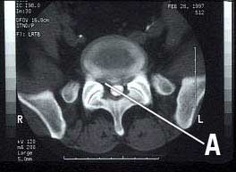
Access to thecal sac [subarachnoid is gained mostly by lumbar puncture. Some authors use C1-C2 puncture. After the access, a water-soluble non-ionic iodinated contrast is pushed into the space. Fluoroscopy-guided lumbar puncture increases the accuracy.
The patient should be rolled several times by rotating and tilting the table to ensure that contrast reaches all the spine.
A CT scan is performed after radiographic contrast has been placed.
After Care
The patient is to lie in bed with the head elevated for 4 to 24 hours after the test. The head is strictly kept elevated so that contrast material does not get into the brain and cause complications.
Strenuous activity should be avoided for at least one day after myelogram. The patient is instructed to report back immediately if there is
- Headache that lasts longer than 24 hours.
- Increase in pain, weakness, or numbness in legs.
- Severe headache, stiff neck, or being sensitive to light
- Bladder or bowel issues
- Fever
- Any other unpleasant occurrence/feeling
Risks & Complications
The complications of CT myelography are mainly because of lumbar puncture and the reaction to contrast material. Most of them are mild but serious complications like allergies are known
Spinal Headache
Spinal headache occurs due to leakage of cerebrospinal fluid after the lumbar puncture, though it is not seen very commonly. The ache begins within 2 to 3 days after the puncture. It becomes worse with sitting and gets better with lying down. It mostly gets resolved by rest and fluid.
Allergic Reaction
Often these are mild and commonly include itching, rash and sneezing. However, though rarely. Serious adverse reactions are rare. A detailed history of allergies may prevent excessive reactions.
Neural Damage
These can occur due to injury due to the needle or bleeding in the space. These may result in weakness or numbness of the treated area. Patients with bleeding disorders are more prone to bleeding complications.
Others
The following are quite rare
- Infection
- Seizures
Results & Inference
Myelogram Normal
- The dye flows evenly through the spinal canal.
- The spinal cord is normal in size, position, and shape. The nerves leaving the spinal cord are normal.
- No narrowing or blockage of the spinal canal is seen.
Myelogram Abnormal
- The flow of dye is blocked or diverted. This may signify compression due to a ruptured (herniated) disc, spinal stenosis, a nerve injury, an abscess, or a tumor.
- Inflammation of the arachnoid membrane.
- One or more nerves leaving the spinal cord are pinched.
References
- Patel D, Weinberg B, Hoch M. CT Myelography: Clinical Indications and Imaging Findings. Radiographics. 2020;40(2):470-84. [link]
- Sandow B & Donnal J. Myelography Complications and Current Practice Patterns. AJR Am J Roentgenol. 2005;185(3):768-71. [Link]