Last Updated on November 13, 2022
Prevertebral space is a space in the neck region defined by the anterior part of the cervical spine and the deep layer of the deep cervical fascia running between the transverse processes of the spine. It is one of the deep neck spaces. There are two others namely retropharyngeal space and danger space.
Anatomy of Prevertebral Space
The prevertebral space is a potential space that lies anterior to the spinal column in the midline.
The space is formed by the alar fascia anteriorly and the prevertebral fascia posteriorly. The prevertebral space extends from the base of the skull to the sacrum/coccyx.
The other two spaces namely retropharyngeal space and danger space that lie in the vicinity [see below] are till mediastinum only.
There is some controversy regarding the content of the prevertebral space. Some authors include these contents within the space whereas others say that these are not to be considered within the space.
The contents are
The space contains
- The prevertebral muscles (longus colli and longus capitis)
- Vertebral vessels
- Scalene muscles
- Phrenic nerve
- The proximal part of the brachial plexus.
Other Neck Spaces of Importance That Need to be Differentiated
There are two spaces that are close by and need to be differentiated from the prevertebral space. These are

Retropharyngeal Space
It extends from the clivus to the upper mediastinum.
It is posterior to the pharynx and esophagus but anterior to the prevertebral muscles.
Its boundaries are formed by
- Buccopharyngeal fascia anteriorly
- Prevertebral fascia posteriorly
- Carotid space laterally
Alar fascia is a part of the deep layer of the deep cervical fascia that extends from the medial border of the carotid space on either side and divides this space into
- Anterior true retropharyngeal space
- Posterior danger space
The true space extends from the clivus inferiorly to a variable level between the T1 and T6 vertebrae.
Danger Space
It extends further inferiorly into the posterior mediastinum to the level of the diaphragm.
It is named so because it forms a passage for the spread of infection from the pharynx to the mediastinum.
Importance of Prevertebral Space
In diseases like osteomyelitis [mostly tubercular], this space can be filled by pus and could be only a radiological sign suggesting the disease.
Prevertebral space may get enlarged due to the pathology of the cervical spine that leads to the accumulation of fluid, blood or pus in the space.
Many a time, enlargement of the space is the finding that raises the doubt about pathology. Infections particularly tuberculosis may otherwise not produce much symptoms and suspicion may be raised from the examination of prevertebral space.
Hematoma accumulation in the prevertebral space can lead to its enlargement following an injury to the neck. Tumors and metastases may spread to this space to cause enlargement.
The conditions that lead to the enlargement of prevertebral space and the consequent increase of its width in x-ray are the following –
- Trauma of the cervical spine
- Vertebral osteomyelitis
- Spondylodiscitis
- Vertebral metastasis.
- Hodgkin lymphoma
- Posterior spread of squamous cell carcinoma head and neck
- Cordoma
- Primary tumors arising from tissues belonging to this space.
Evaluation
The PV space needs to be evaluated radiologically. It can be done by x-ray, CT or MRI.
X-ray
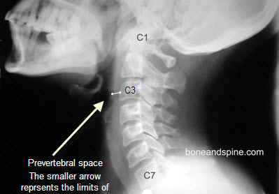
This space is evaluated on the lateral view of the cervical spine as seen above. It is one of the important components of the cervical spine x-ray evaluation.
On lateral cervical spine x-ray, the normal prevertebral space looks like this.

The normal dimension in the adult is 4 mm at C3 level. A greater than 7 mm value is definitely abnormal and the cause should be looked for.
If the values lie in between, a clinical correlation must be sought.If space is increased a computed tomogram or MRI should be done for further evaluation of pathology.
At C6/7 levels, the normal value is less than 21 mm.
CT
CT provides greater details of bone destruction and can identify the lesions with fair certainty though MRI does a better job.
The various normal values for PV space on CT are
C1: 8.5 mm
C2: 6 mm
C3: 7 mm
C4/C5: variable
C6/7: 18 mm
The values for children differ due to the presence of adenoid tissue. The values are different for different age groups.
MRI
MRI is the preferred investigation in these cases. It not only identifies the soft tissue with better details, it also identifies various disease processes. For example, it can detect tuberculosis much earlier than x-ray.
References
- Branstetter BF, 4th, Weissman JL. Normal anatomy of the neck with CT and MR imaging correlation. Radiol Clin North Am. 2000;38(5):925–40.
- Davis WL, Harnsberger HR. CT and MRI of the normal and diseased perivertebral space. Neuroradiology. 1995;37(5):388–94.
- Silver AJ, Mawad ME, Hilal SK, et al. Computed tomography of the nasopharynx and related spaces. Part II: pathology. Radiology. 1983;147(3):733–8.