Last Updated on November 22, 2023
The wrist is a complex joint formed by a collection of multiple bones and joints bridges the hand to the forearm. It is an ellipsoidal joint.
The bones comprising the wrist include
Wrist carries complex articulations that allow variable mobility of the hand. Relative to the forearm, the hand is capable of 3 degrees of freedom.
- Flexing and extending
- Pronation and supination
- Ulnar and radial deviation
The wrist is well stabilized by its ligaments without sacrificing the stability of the joint.
Bones of the Wrist
As noted, eight carpal bones, the distal radius and ulnar, and the bases of the 5 metacarpals participate in the wrist joint. The ulna is not part of the wrist joint – it articulates with the radius, just proximal to the wrist joint, at the distal radioulnar joint. It is prevented from articulating with the carpal bones by a fibrocartilaginous ligament, called the articular disk, which lies over the superior surface of the ulna.
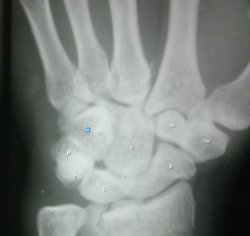
There are eight carpal bones arranged in two rows. Together, the carpal bones form a convex surface, which articulates with the concave surface of the radius and articular disc.
[Read more on carpal bones anatomy]
The proximal row is found at the level of the distal wrist crease and includes the scaphoid, lunate, triquetrum, and pisiform from radial to ulnarward. The second row of carpal bones, the distal row, is made up of the trapezium, trapezoid, capitate, and hamate.
The bones in the proximal rows articulate with distal ends of radius and ulna on one side and bones in the distal carpal rows on the other.
The distal carpal row articulates with bases of five metacarpals.
The proximal carpal row bones represent an intercalated segment, as no tendons insert upon them.
The movement of this row, therefore, is dependent entirely on mechanical forces from surrounding joints.
The bones of the distal row are closely adherent to each other via intercarpal ligaments as well as tightly bound to the metacarpal bones by carpometacarpal joint.
The ligamentous connection between the trapezoid and capitate to the index (second) and middle (third) finger metacarpals, respectively, are so rigid that the distal carpal row has been considered a component of a fixed hand unit that moves in response to musculotendinous forces generated from the forearm.
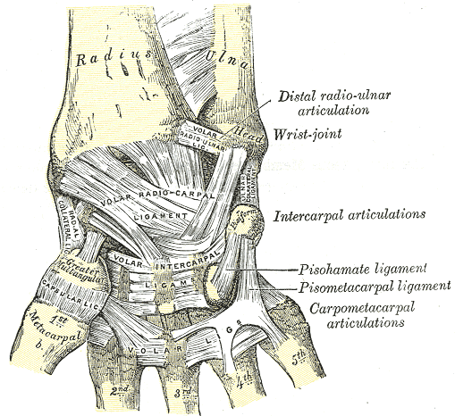
Joint Capsule
The capsule is double layered. The fibrous outer layer is attached to the radius, ulna and the proximal row of the carpal bones. The internal layer is comprised of a synovial membrane, secreting synovial fluid which lubricates the joint.
Ligaments of Wrist
The wrist has multiple ligaments that provide stability.
Superficial ligaments on the volar side are
- Palmar aponeurosis
- Radial collateral ligament
- Triangular Fibrocartilage complex
- Ulnar Collateral Ligament
- Flexor Retinaculum
Extensor Retinaculum is the main superficial ligament on the dorsal side.
These are the main stabilizing ligaments and include extrinsic and intrinsic ligaments and carpometacarpal ligaments.
Different patterns of naming ligaments of the wrist can cause confusion. Therefore, it is better to remember them by the bones they bind together.
The ligaments can be divided into two categories palmar or volar and dorsal.
Another division of the ligaments is extrinsic and intrinsic.
Extrinsic ligaments are the ligaments that link the carpal bones to the radius, ulna, and metacarpals. these ligaments are attached to roughened areas on the dorsal and palmar surfaces of the carpal bones.
Intrinsic ligaments are the ligaments that connect the carpal bones to each other.
Ligaments of the wrist are most highly developed on the palmar side of the wrist as compared to the dorsal side.
Extrinsic Ligaments of Wrist
Palmar Radiocarpal Ligaments
The palmar radiocarpal ligaments originate from the distal palmar margin of the radius and extend to the carpus. They are stout structures and the primary stabilizes of the radiocarpal joint. Although when viewed from outside the joint they appear as a confluent sheet of the capsule, these ligaments are distinguishable arthroscopically.
Radial Collateral Ligament
The radial collateral ligament, located on the thumb side of the wrist, attaches the distal end of the radius to the trapezium bone as well as the scaphoid bone. This ligament restricts ulnar deviation.
[On the ulnar side, there is another collateral ligament, which attaches the distal end of the ulna to the triquetral
bone and the pisiform bone. It restricts and restricts radial deviation]
Radioscaphocapitate Ligament
It is located ulnar to the radial collateral ligament.
It originates from the radial styloid and proceeds in an ulnar-oblique direction with various insertions on the waist and distal pole of the scaphoid before terminating on the neck of the capitates. It forms the radial arm, while the ulnocapitate ligament [see below] forms the ulnar arm [termed the arcuate ligament complex].
The radioscaphocapitate ligament serves as a fulcrum around which the scaphoid flexes.
An excessive radial styloidectomy may jeopardize its origin, resulting in abnormal scaphoid motion.
For preserving, a short oblique osteotomy with the removal of only a few millimeters of articular surface should be done.
The radioscaphocapitate ligament should be preserved when performing a proximal row carpectomy, as it will be the only remaining palmar radiocarpal ligament.
Long Radiolunate Ligament
It originates at the distal radius from the volar aspect of scaphoid fossa where the attachment is partially overlapped by the radioscaphocapitate ligament. It lies ulnar to radioscaphocapitate ligament.
It was previously called the radiolunotriquetral ligament. The long radiolunate and radioscaphocapitate ligaments are separated by an interligamentous sulcus, which is site us is the site of the palmar arthroscopic portal.
It inserts radially on the distal surface of the lunate as well as the volar segment of the scapholunate interosseous ligament. It does not directly attach to the scaphoid.
The long radiolunate ligament provides stability to counteract ulnar or distal translocation of the lunate. It is also called radiolunotriquetral or volar radiolunate ligament and counters ulnar-distal translocation of the lunate. It is abnormal in Madelung’s deformity
Radioscapholunate Ligament
It arises from the ridge between the scaphoid and lunate facets and merges into the proximal membranous portion of the scapholunate interosseous ligament.
It is also known as the ligament of Testut and passes along the ulnar side of the long radiolunate ligament. Although usually called a ligament, it functions primarily as a conduit for terminal vessels and nerves from the radial artery and anterior interosseous neurovascular bundle.
This ligament, therefore, does not add to the mechanical strength.
Short Radiolunate Ligament
The short radiolunate ligament is the most ulnar of the radiocarpal ligaments and is separated from the long radiolunate ligament by the radioscaphocapitate ligament. It originates from the palmar rim of the lunate fossa and inserts on the proximal radial edge of the plamar horn of the lunate.
It along with other radiocarpal ligaments resists the natural tendency of the carpus to translate. Posttraumatic ulnar translation indicates extensive injury to these ligaments.
The short radiolunate ligament originates at the distal radius on the ulnar and volar edge of the lunate fossa and inserts into the volar aspect of the lunate. Its insertion is located proximal to the insertion of the ulnolunate ligament. The short radiolunate ligament has a potential role in lunate stabilization.
Palmar [Volar] Ulnocarpal ligaments
Three confluent ligaments form the ulnocarpal ligament complex.
The palmar ulnolunate and ulnotriquetral ligaments arise from the palmar radioulanar ligaments, which is a component of the triangular fibrocartilage complex, and insert into their respectively named carpal bones. Ulnocapiate is the third palmar ligament.
The palmar ulnocarpal ligaments stabilize the ulnocarpal joint, and their injury may result in a supination deformity of the carpus, often called an ulnar sag [in rheumatoid arthritis, trauma].
Avulsion fractures of the TFCC from its ulnar attachment may cause direct injury to the ulnocapitate and ligament and indirect injury to the other two ligaments.
Ulnolunate ligament
It originates radial to the ulnocapitate ligament at the base of the ulnar styloid process. It inserts on the ulnar portion of the palmar horn of the lunate, distal to the insertion of the short radiolunate ligament and becomes confluent with it.
Ulnotriquetral Ligament
This ligament passes to the ulnar lobe of the triquetrum, where it forms the distal portion of the extensor carpi ulnaris subset. The ulnotriquetral ligament commonly has a small distal rent that creates communication between the radiocarpal and pisotriquetral joints.
Ulnocapitate Ligament
The ulnocapitate ligament originates from the fovea of the ulnar head and the base of the ulnar styloid and lies just palmar to the ulnolunate and ulnotriquetral ligaments to insert on the neck of the capitates. It forms the ulnar arm of the arcuate ligament complex [radioscaphocapitate ligament forms the radial arm]
It distally inserts onto the proximal and volar region of capitate.
Space of Poirier
Just proximal to the apex of the arcuate ligament complex [ ie, between the distal portions of the ulnocapitate and radioscaphocapitate ligaments], the palmar wrist capsule is devoid of ligaments.
This relatively weaker region of the capsule is called the space of Poirier.
This is the region where the mid carpal joint dislocates in a perilunate dislocation.
With palmar flexion of the wrist, an area of weakness disappears and in wrist dorsiflexion, the area of weakness increases
In perilunate dislocations, this space allows the distal carpal row to separate from the lunate.
In lunate dislocations, the lunate escapes into this space.

Dorsal Extrinsic
Dorsal Intercarpal Ligaments (DIC)
The dorsal intercarpal ligament originates from the dorsum of the triquetrum and inserts on the dorsal aspects of the trapezoid and the waist of the scaphoid.
Dorsal Radiocarpal Ligament is the collective term for the following three ligaments
- Radiolunate
- radioscaphoid
- Radiotriquetral*
Few slips go to capitate as well.
Along with the radiocarpal ligament, the intercarpal ligament maintains carpal stability and alignment while preventing dorsal intercalated segment instability (DISI) and volar intercalated segment instability (VISI) deformities.
* Radiotriquetral ligament must also be disrupted for VISI deformity to form (in combination with rupture of lunotriquetral interosseous ligament rupture.)

Intrinsic Ligaments of Wrist

These are the ligaments which span from one carpal bone to other. Some authors further differentiate them into interosseous and capsular ligaments but for the sake of simplicity, I have kept them together.
Basically, Interosseous ligaments lack a fibrous lamina and are located entirely within the joint.
The terms interosseous and capsular are also more consistent with clinical practice, where the term interosseous ligaments are typically used to describe the scapholunate and lunotriquetral ligaments, which are often the focus of a wrist injury evaluation.
Row-wise ligaments are described below
Intrinsic Ligaments of Proximal row
Scapholunate ligament
The scapholunate ligament connects the scaphoid and lunate along the proximal joint surface and is the primary stabilizer of the scapholunate joint.
The ligament has three parts – volar portion, dorsal portion and a proximal part. The dorsal portion is the thickest of the most critical to prevent translation.
Volar portion is important to prevent rotation.
The proximal portion has got no significant strength.
Disruption of this ligament allows the lunate to extend when the scaphoid is flexed, creating a dorsal intercalated segment instability (DISI) deformity.
Lunotriquetral Ligament
The lunotriquetral ligament attaches the lunate and triquetrum along the proximal joint surface.
It also has a dorsal, volar and proximal portion.
Injury to the lunotriquetral ligament permits lunate flexion with a normally aligned scaphoid, creating a volar intercalated segment instability (VISI) deformity (see the image above)
The lunotriquetral ligament originates from the ulnar side of the lunate and attaches to the triquetrum distal to the attachment of the ulnolunate ligament and radial to the lunotriquetral interosseous ligament.
The disruptions of this ligament lead to lunate flexion when the scaphoid is normally aligned creating VISI deformity (in combination with rupture of dorsal radiotriquetral rupture)
Distal Row Intrinsic Ligament
Trapeziotrapezoid Ligament
The trapeziotrapezoid ligament joins the trapezium and trapezoid and lies entirely within the trapeziotrapezoid joint.
It attaches to the volar side of the ulnar aspect of the trapezium and travels ulnarly to the distal radial edge of the trapezoid.
Capitotrapezoid Ligament
The capitotrapezoid ligament attaches the capitate to the trapezoid. This ligament travels from the central region of the ulnar side of the trapezoid to the distal, radial side of the capitate.
Capitohamate Ligament
The capitohamate ligament is composed of distal and proximal ligaments.
The distal component of the capitohamate ligament originates on the distal side of the ulnar edge of the capitate and attaches to the distal side of the radial edge of the hamate.
The proximal ligament originates on the ulnar edge of the capitate and attaches to the proximal side of the radial edge of the hamate.
Palmar midcarpal Ligaments
The palmar midcarpal ligaments are short, stout capsular ligaments that cross a single articulation.
The triquetrocapitate and triquetrohamate ligaments blend with the distal fibers of the UC ligament and reinforce the ulnar arm of the arcuate ligament. These ligaments are stabilizers of the scaphoid, triquetrum, and midcarpal joint.
Scaphotrapeziotrapezoid Ligament
Scaphotrapeziotrapezoid ligament passes from the palmar surface of the distal pole of the scaphoid to the palmar surface of the trapezium and trapezoid. Some consider the two as different ligaments.
This ligament is composed of a radial branch and an ulnar branch. Both of them attach to scaphoid tuberosity.. Branches diverge, with the radial branch attaching to the radial aspect of the trapezium, whereas the ulnar branch attaches to the proximal side.
Scaphocapitate Ligament
The scaphocapitate ligament originates from the distal pole of the scaphoid and inserts on the body of the capitates. It lies parallel to the radioscaphocapitate ligament.
Triquetralcapitate Ligament
The triquetrocapitate ligament attaches proximally to the volar and radial edge of the triquetrum and inserts distal to the ulnar and volar edge of the capitates.
Triquetralhamate Ligament
The triquetrohamate ligament proximally attaches to the distal edge of the triquetrum and distally to the dorsal edge of the hamate. The triquetrocapitate ligament may overlap the triquetrohamate ligament
Anatomical Variations of Wrist
The shape of the fourth metacarpal base may vary from being broad-based to narrow-based.
Ossification and shape of lunate may vary [medial facet may or may not be present]
Distal articulation of the scaphoid with trapezium may have separate facets or combined facet.
Isolated carpal coalition [lunotriquetral, capitohamate] may occur.
Neurovascular Supply of Wrist
The wrist joint receives blood from branches of the dorsal and palmar carpal arches, which are derived from the ulnar and radial arteries.
Nerve Supply of Wrist
Following branches supply the wrist joint
- Median nerve via the anterior interosseous branch.
- Radial nerve via posterior interosseous branch.
- Ulnar nerve via deep and dorsal branches.
Movements of the Wrist Joint
The wrist is an ellipsoid type synovial joint, allowing for movement along two axes. This means that flexion, extension, adduction, and abduction can all occur at the wrist joint.
All the movements of the wrist are performed by the muscles of the forearm.
The wrist motion is as a result of complex arrangements between the two carpal rows. Wrist’s versatility mostly is due to the intercalated three-bone system of the proximal carpal row.
Flexion
In this movement, the hand and wrist is moved toward the palmar surface of the forearm
It is produced mainly by the flexor carpi ulnaris, flexor carpi radialis, with assistance from the flexor digitorum superficialis.
Extension
When the hand and wrist are moved towards the dorsal surface of the forearm.
It is produced mainly by the extensor carpi radialis longus and brevis, and extensor carpi ulnaris, with assistance from the extensor digitorum.
Adduction or Ulnar Deviation
The hand deviates towards the radial side.
It is produced by the extensor carpi ulnaris and flexor carpi ulnaris
Abduction or Radial Deviation
The hand deviates towards radial side
It is produced by the abductor pollicis longus, flexor carpi radialis, extensor carpi radialis longus, and brevis.
Axial Rotation
There is another movement occurring at the distal radio-ulnar joint which is the axial rotation. This movement enables supination and pronation.
Carpal bones contribute about 10 degrees of rotation while rest is contributed by forearm [about 140 degrees arc]
Normal wrist motion
- Flexion and extension – 65-70 degrees
- Radial deviation – 20 degrees
- Ulnar deviation is 40 degrees.
Mechanics of Motion
Flexion/Extension
During flexion and extension, each carpal row angulates to an equal extent in the same direction.
Radioulnar Deviation
For radioulnar deviation, the proximal row undergoes a secondary angulation in the sagittal plane [A plane that divides the body into left and right equal halves] in addition to the synchronous motion in the coronal plane.
During radial deviation scaphoid undergoes flexion as the trapezium comes towards the radius. Scapholunate ligament transmits this motion the lunate and triquetrum.
With movement in opposite direction towards neutral and further to the ulnar deviation, the proximal row extends and supinates.
With ulnar deviation, scaphoid extends. Proximal migration of the hamate forces the triquetrum to displace palmarly and extend and lunate comes along. Following video shows the movement of carpal bones under fluoroscopy.
Clinical Significance of Anatomy of Wrist
Wrist Instability
By varying the length and contour, the proximal carpal row permits extensive excursion of the wrist and at the same time maintains stability.
Disruption of the mechanism due to a fracture or ligamentous injury results in instability of the wrist due to loss of synchronicity and change in contact pattern of carpal bones.
Instability of the wrist, over time, result in arthritis of the wrist.
Injuries to the Wrist Joint
Fracture of the Scaphoid
Scaphoid is a common carpal bone to fracture and often breaks when a fall occurs on an outstretched hand.
Because the scaphoid has a unique blood supply, it is at risk to undergo necrosis after the injury.
The main clinical sign of a scaphoid fracture is tenderness in the anatomical snuffbox.
Anterior Dislocation of the Lunate
This usually occurs by falling on a dorsiflexed wrist. The lunate is forced anteriorly, and compresses the carpal tunnel, causing the symptoms of carpal tunnel syndrome.
Other Injuries
Fractures of the distal radius, distal ulna, and other carpal bones also occur.