Last Updated on December 19, 2019
Elbow injuries are very common in adults and children. The treatment of injuries depend on the pattern of injury and age of the patient along with many other factors.
Following are various xrays of elbow injuries in adults and children
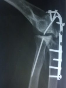
Image 1 – Xray of Operated Fracture Lateral Condyle in a Child

The image today is an x-ray of operated fracture lateral condyle, done immediately after surgery. As viewed, the xray shows a fracture of lateral condyle which is held in position by a Kirschner wire and a screw.
The K-wire is longer than it should be. A longer protruding implant beyond the bone is dangerous for nerves and vessels.
Image 2 – Xray of Fracture Lateral Condyle of Humerus
The Xray in the picture shows a fracture lateral condyle of the humerus.

This x-ray belongs to fourteen years old male who hit his elbow when he fell from a cycle.
Image 3 – Xray of Posterior Dislocation of Elbow

The image shows a posterior dislocation of the elbow. This is an x-ray of a 60 years old woman who suffered household trauma but did not take any treatment for 4 weeks. There are some bone fragments visible too.
All dislocations are emergency. This elbow was neglected for 4 weeks and could not be reduced with closed methods. It was operated and functional outcome is acceptable.
To avoid all kind of complications, all dislocations should be reduced immediately. But the first step in that is to seek medical help.
Image 4 – Fracture Of Lateral Condyle of Humerus In A Child – Anteroposterior and Lateral Views
Fracture of lateral condyle is a very common fracture in the children. Following x-ray is of an eleven-year-old boy who fell down while playing.
 On the left-hand side is the lateral view which also shows dislocation of elbow whereas on the right-hand side is anteroposterior view.
The boy was treated with open reduction and internal fixation using three k-wires.
Image 5 – Dislocation of Elbow With Fracture of Radial Neck – AP and Lateral Views
45 years old lady came to the outpatient department with a history of fall from two wheeler 3 days back and complained of swelling in the left elbow and inability to use the limb.
On examination, there was tenderness around the elbow and any movement elicited the pain. An x-ray of the elbow that she had carried along revealed following picture.

Though the x-ray is not of good quality, it is able to show the dislocation and radial neck fracture.
The patient was offered treatment by closed reduction of the elbow and if required open reduction and fixation of radial neck.
Patient due to some personal reasons refused the treatment. She did not visit in follow up also.
Image 6 -Xray Of Fracture 0f Medial Epicondyle Of Humerus In 14 Years Old Girl
The following x-ray depicts an anteroposterior view of the elbow of 14 years old girl who fell from a mango tree. She presented with pain and swelling of the elbow.
The x-ray looked like this, showing a fracture of medial epicondyle of the humerus.
She did not have any ulnar nerve deficit. The elbow was not grossly unstable.
She was managed in above elbow plaster cast.
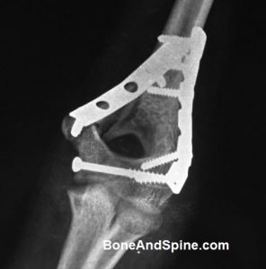
Image 7 – Xray of United Fracture of Lateral Condyle Of Humerus
Following is the 8 weeks postoperative x-ray showing union of fracture in lateral condyle of the humerus. 
The preoperative photograph is here.
Image 8 – Anteroposterior Xray Of Elbow of a Child Showing Fracture Of Neck Of Radius
Fractures of the neck of the radius are common in children as compared to radial head fractures which are commoner in adults.
Following xray is of 11 years old child witha fracture of radial neck. AS the xray quality is not good, I have encircled the area of interest.

The fracture was managed by closed manipulation and above elbow cast.
Image 9 – Monteggia Fracture Dislocation Xrays
Monteggia fracture involves the fracture of shaft of ulna and dislocation of the radius. Following xray is of 39 years old male who suffered a motor vehicle injury to his upper limb and found to have a Monteggia fracture.

As per Monteggia fracture classification, the fracture is Bado type I.
The fracture was treated by open reduction and internal fixation using low contact dynamic plate and transfixing the radial head to the ulna.
Image 10 – Postoperative Film of Supracondylar Fracture of Humerus In 16 Years Old
Following is the postopeartive xrays of supracondylar fracture of humerus shown here.

The fracture was approached by the posterior approach and fixed with two reconstruction plates.
Image 11 – Xray Of Supracondylar Fracture In A 16 Year old Child
The lateral view reveals supracondylar fracture very well and also bent kwires from a previous surgery for fracture lateral condyle.

The anteroposterior xray of the elbow is of quite poor quality. You would need to strain to see the fracture line. Unfortunately, there was only this preoperative image available.

The supracondylar fracture was treated by open reduction and internal fixation with reconstruction plates and distal epiphyseal injury to radius was treated with closed reduction and Kwire fixation.
Image 12 – Xray of Elbow Showing Fracture Of Capitellum
Fracture of capitellum in 43 yars old lady. 
She refused for any treatment though.
Image 13 – Neglected Dislocation Of Elbow In A Child
Xray of negelcted elbow dislocation in 10 years old child.

There is a periosteal reaction present on the posterior side.
Image 14 – Xray Of Intercondylar Fracture of Humerus With Diaphyseal Extension
Anteroposterior and lateral view xrays of a young adult with fracture of distal humerus extending up to diaphysis.

The patient was treated with open reduction and internal fixation using long recon plates and cancellous screws.
Image 15 – An Unusual Fracture Of Proximal End Ulna Along With Dislocation of Radial Head
An unusual of fracture of upper end of the ulna

The fracture involves a vertical split upper part of the shaft of ulna along with the coronoid process of the ulna.
There is also dislocation of the radial head.
Image 16 – Lateral View Radiograph of Fracture Capitellum
Lateral Xray of Elbow showing Capitellum fracture

A smaller comminuted fragment is visible as well
Image 17 – Xray Elbow Showing Old Ununited Fracture Lateral Condyle With Cubitus Valgus
An ununited fracture of the lateral condyle can lead to cubitus valgus as the growth occurs.
Following is an xray of an adult who had suffered a fracture of lateral condyle in childhood and was not treated for the same

The xray shows an united fracture of lateral condyle and elbow in a high degree of valgus.
Image 18 -AP and Lateral Xrays of Elbow Showing Comminuted Supracondylar Fracture of Humerus
33 years old lady with a history of motor vehicle accident suffered an injury to her right elbow and xray of the elbow revealed a supracondylar fracture.

The fracture was grade I open fracture.
Image 19 -Fracture Of Radial Head and Medial Epicondyle Of Humerus
Xray of the elbow of 13 years old boy after injury.

The injury shows a fracture of the radial head with a fracture of the ipsilateral medial condyle of the humerus.
The injury resulted due to fall from a bicycle.
Image 20 -Xray Elbow Showing Nonunion of Fracture Lateral Condyle Humerus With Cubitus Valgus
Cubitus Vlagus is the deformity of the elbow resulting in forearm pointing outwards in relation to the arm. One of the late complication of nonunion of humerus lateral condyle fracture is cubitus valgus.
Following is an xray of an adult who presented with deformity of cubitus valgus and xray revealed non union of the lateral condyle.

He gave a history of childhood injury for which no treatment was taken.
Image 21 – Xrays of Radial Neck Fracture In A Child
AP and lateral views of elbow showing nadial neck fracture in a child

Fracture has been marked with arrows in both the views.
AP and Lateral Views Of Undisplaced Fracture of Neck of Radius
Image 22 – Fracture of the radial neck in an adult.
AP view

Lateral View

The fracture is undisplaced and has been marked with an arrow.
Image 23 -Xray of Elbow Showing Fracture of Head of Radius
Xray of elbow showing a fracture of head of the radius in 39 year old male

The fracture has been marked with an arrow.
Image 24 – Supracondylar Fracture of Humerus With Fracture of Medial Epicondyle
Four years old child with a history of fall on elbow showing a fracture in supracondylar part of the elbow and medial apicondyle

Both fractures have been marked with an arrow.
Image 25 – Fracture Dislocation of Elbow

Anteroposterior and lateral view of xray of 35 years old male showing a fracture of olecranon extending till the upper fourth of the ulnar shaft, fracture of cornoid, dislocation of ulno humeral joint and radial head.
A very severe trauma is required to produce this kind of injury in an adult. This patient suffered an injury in motor vehicle injury.
Image 26 – Xray Showing New Bone Formation In Excised Radial Head Leading To Ankylosis
After achieving a good initial movements for 2 months, this 40 years old female, a follow up case of radial head excision started having stiffness in supination and pronation movements of the affected elbow.
The following xray was done after 3 months of recurrence of symptoms.

It shows new bone formation in the excision area which resulted in ankylosis.
Image 27 – Xray of Fracture of Medial Epicondyle of Humerus and Ipsilateral Radial Head
Xray of elbow showing a fracture of medial epicondyle and radial head.

The other view of the elbow is not available.
Image 28 – Postoperative Xray of Elbow After Radial Head Excision
Postoperative xray of the elbow after radial head excision done for radial head fracture.

Staples used in suturing the incision can also be seen.
Image 29 – Fracture Through Elbow With Previously Bony Ankylosis and Flexion Deformity
43 years old male patient fell from the bike and injured his elbow. The xray revealed a fracture in the ankylosed elbow. The ankylosis was bony type in nature.
The patient could not provide reliable history or documents related to ankylosis.

The patient was treated in fiber cast after positioning the limb in the same amount of deformity.
The xray above was taken after 4 weeks of application of cast and shows a uniting fracture.
The fracture united quite well eventually.
Image 30 -Xray Elbow Showing Intercondylar Fracture of Humerus
Xray of fracture of the intercondylar region of the humerus.

The fracture occurred following a motor vehicle injury and was an open fracture.
Image 31 – Xray of Dislocation of Elbow Joint
Xray of dislocation of the elbow in 19 years old male.

The dislocation was treated by closed reduction and above elbow plaster slab.
Image 32 – Xray Elbow Showing Ulna United In Malposition and Ununited Radial Head Fracture
Forty eight years old lady came to OPD after two months of injury with a complaint of the stiffness of elbow. She had been treated conservatively at some other hospital.
Previous xrays revealed a fracture of radial head and fracture of upper end of the ulna.
Her extremity was wasted as compared to opposite limb and had an angular deformity.
A frash xray was done and revealed malunited ulna and ununited radial head fracture.

She was advised surgical correction of the deformity and excision of radial head. She refused.
Image 33 – Clinical Photograph of Elbow Dislocation

Note the prominent olecranon which appears more posterior than its usual position and a taut prominent triceps. The elbow is kept in attitude of flexion.
The xray revealed a posterior dislocation of elbow.
The dislocation was reduced and an above elbow plaster was applied in flexion.
Image 34 – Xray of Elbow With Ununited Olecranon and Broken Implant
Following xray is of an elbow which must have been operated for fracture of the olecranon and radial head dislocation [asumption- the original history not available]
The xray shows a broken plate that fixed olecranon and a broken kwire that passes from humerus to radius.
Image 35 – Xray of Reduced Monteggia Fracture Dislocation In A Child With Ulna In Malposition
Nine years old child fell from the height and had Monteggia Fracture Dislocation [ Monteggia fracture dislocation involves fracture of upper ulna bone and dislocation of the radial head from the elbow.]
The fracture was reduced and above elbow plaster was applied.

The xray above is taken after application of plaster and shows a reduced radial head but ulna is in malposition. The patient was advised surgery for correction of the position but patient guardians refused.
Image 36 – Xrays of Supracondylar Fracture of Humerus Before And After Surgery
53 years old male fell from the scooter and injured his elbow. An xray of the elbow revealed a supracondylar fracture of the humerus.
The xray is shown below

The fracture was treated by open reduction and internal fixation using reconstruction plates. Following image was taken just after the surgery using C arm image intensifier.

Patient is still under follow up and showing good improvement.
Image 37 – Xray of Elbow Showing Fracture of Head of Radius
Xray of elbow showing a fracture of head of the radius in 39 year old male
The fracture has been marked with an arrow.
Image 38 – deleted
Image 39 -Xrays Intercondylar Fracture of Humerus Treated By Open Reduction and Internal Fixation
Following xrays are of 15 years old male who had intercondylar fracture of humerus. Here is AP view of the elbow showing intercondylar fracture of humerus.

Same fracture on lateral view x-ray.

The fractures were fixed using screws and plates. Here is the AP view after fixation
Lateral view

The boy regained almost full range of motion of the elbow.
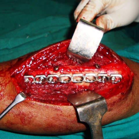
Image 40 – Photograph Excised Radial Head Reconstructed on Trolley
Radial head excision procedure is done for those injuries of the radial head where open reduction and internal fixation is not an option. This may be either due to the severity of the injury itself, low patient demands or the patient’s decision to remove the radial head instead of preserving it.
After removal of the radial head, it is important to ensure completeness of the removal by reconstructing the radial head preoperatively.

This photograph is of a removed and reconstructed radial head.
You can visualize the complete radial head.
Image 41 – Fracture Upper-End Ulna Fixed with Reconstruction Plate and Interfragmentary Fragment
Following x-rays are of fracture of upper end of the ulna and belong to a laborer who fell from a height.
Here is AP view

Lateral view

The fracture was treated by open reduction and internal fixation using interfragmentary screws and reconstruction plate.
Here is peroperative photograph taken during surgery.
The fracture united within 6 weeks and the patient achieved an almost full range of motion of the elbow.
Image 41 – Minimally Displaced Fracture of Olecranon
Fracture of olecranon in 45 years old male who fell from a height. The fracture had minimum displaced.

The fracture was treated by a plaster cast.
Image 42 – Xray of Fracture of Supracondylar Region of Humerus
Fracture of the supracondylar region of the humerus in 39 years old female who suffered this injury in a motor vehicle accident.
AP view
Lateral view

There was no neurovascular deficit. The patient was treated with open reduction and internal fixation using reconstruction plates.
Image 43 – Xrays of Fracture of Radial Neck In Adult
Fractures of the radial neck are relatively uncommon injury around the elbow in the adults. This injury is more common in children. In contrast, radial head fractures are more common in adults.

The x-ray in the image is of 48 years old lady who fell from a scooter. This fracture generally results from a hard fall on an extended & supinated outstretched hand. The force is transmitted through the shaft of radius and momentum of body drives capitellum (A projection of lower humerus that articulates with head of the radius) against lateral half of radial head, tilting & displacing it laterally.
- If the forearm is fully supinated, the displacement is lateral
- If the forearm is in neutral mid position, displacement is posterior
In the present x-ray a lateral displacement is noted.
Image 44 – Fracture Of Radial Head – Anteroposterior View of Elbow
Radial head fractures occur in isolation or in association with fracture-dislocations around the elbow. The following x-ray is of 37 years old male who suffered trauma to his right elbow following a fall in a pit.
The patient presented to us about 3 weeks after the injury and was treated by radial head excision.
It is worthy of noting that had this patient presented earlier, there were strong chances of radial head preservation.
The treatment depends upon the displacement of the fracture. A mildly displaced fracture like in the picture may be amenable to closed reduction but a severe displacement may warrant open reduction and internal fixation.
The lady refused any treatment for this and was sent home in a splint.
Image 45 -Comminuted Fracture Of Olecranon Process Of Ulna
This x-ray was brought to the OPD for an opinion. The patient was an 85-year-old lady who was struck by a motorbike and fell down.
The x-ray showed a comminuted fracture of olecranon that consisted of two large fragments, separated from ulna and from each other.

The person was advised to bring the patient so that she could be assessed but never showed up.
Image 46 – Anteroposterior and Lateral Xrays of Elbow Showing Fracture of Radial Head
Fracture of the radial head in a 38-year-old female.

The anteroposterior view above shows a fracture of the radial head.

The lateral view shows comminution as well.
Image 47 – Xray of Fracture Lateral Condyle In Two Years Old Child
Fracture of lateral condyle is very common fracture in children. lateral condyle is part of the humerus bone that projects on outer aspect of lower part of the arm just above the level of flexor crease of the elbow (cubital fossa).

Most of the undisplaced fractures can be managed conservatively. The surgery is required in cases which have the displacement of the fragment more than 2mm or the fragment is rotated.
As this is a type of epiphyseal injury, the anatomical reduction is absolutely necessary. Otherwise, growth disturbances of elbow and deformity of the elbow may result, most common being cubitus valgus or valgus deformity of the elbow.
The x-ray above is of two years old child who sustained an injury due to fall from a bed. The fracture is not displaced and was managed conservatively.
Image 48 – Xray of Flexion Type Supracondylar Fracture In A Child
This is an x-ray of a five-year-old boy who sustained an injury to the elbow after fall. The anteroposterior and lateral view of elbow [see image] revealed a flexion type supracondylar fracture.
Flexion type supracondylar injuries are rarer as compared to extension type

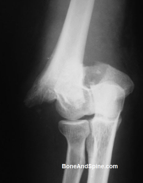
Image 49 – Dislocation of Elbow in a Child
Dislocation of the elbow in children is less common than an adult. Here is an x-ray of elbow lateral view in 12 years old child.

The patient was treated with closed reduction of the dislocation and plaster splint.
Image 49 – Nonunion of Fracture Lateral Condyle of Humerus

Image 50 – Fracture of Radial Neck in a Child
The following x-ray shows a fracture of a radial neck in a child.

The mode of injury is not known.
[amazon_link asins=’B00O66AJOG,B079B8FR27,B01G770J6E,B01H2123YU,B000KGOMBC,B00QY0EFJM’ template=’CopyOf-ProductCarousel’ store=’bas0ba-2b’ marketplace=’US’ link_id=’66aa684c-fc7f-11e8-9754-93a8550f4ee0′]