Last Updated on July 31, 2019
Another term for ingrown nail is onychocryptosis which literally means hidden nail. It is also known as unguis incarnates. Ingrown nail is a condition where the nail grows to cut into one or both sides of the paronychium or normal toenail entering in the skin that is overgrown, mostly precipitated by an infection.
Causes of Ingrown Toenail
The main cause of ingrown toenail are
- Ill-fitting footwear especially those with inadequate toe-box room
- Damp atmosphere of enclosed shoes, softening the nail-plate
- Swelling the epidermal keratin
- Improper cutting of the nail cutting the nail- too short, rounded off at the tip or peeled off at the edges
- Trauma to the nail plate or toe
Pathophysiology
Ingrown toenail is rare in people who do not wear shoes. The condition is resulted by a combination of factors like an improperly trimmed nail and ill footwear. When a shoe is worn, the great toe is pushed toward the second toe and this results in pressure against the lateral border of the nail. This pushes the nail fold into the sharp edge of an improperly cut nail. Infections occur in the open wound resulting in inflammation leading to abscess which drains poorly.
Granulation tissue forms which is slowly covered by epithelium, further inhibiting drainage and promoting edema.
The nail becomes vulnerable to injury by extrinsic pressure, and the cycle continues.
Risk Factors for Ingrown Toenail
Few people and conditions are said to be prone to ingrown toenails. These include tight or narrow shoes, repeated injury to feet, poor hygiene of the feet, poor posture and gait, congenital foot deformities, long toes, naturally short nails, obesity, diabetes, infections, prior nail surgery, and excessive foot sweating increase the risk of ingrown toenail.
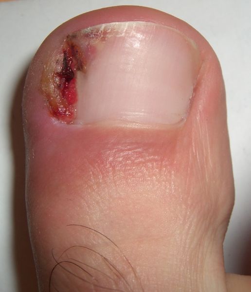
Stages of Ingrown Toenail
Stage I (Inflammatory Stage)
The patient has mild erythema [redness], swelling, and tenderness along the lateral nail fold.
Stage II (Abscess Stage)
The erythema, edema, hyperhidrosis [sweating] and tenderness increase. The lateral nail fold bulges over the lateral nail plate edge, and drainage begins. Infection follows, and initial serous drainage becomes purulent. Walking and shoe wear is almost impossible.
Stage III (Granulation Stage)
Granulation tissue covers the lateral nail fold and inhibits free drainage. I untreated cases, epithelium covers the edge of the granulations causing drainage block. This can progress into a chronic condition occasionally exacerbated by acute episodes
Diagnosis
The diagnosis is clinical and usually, imaging is not required except for the advanced cases where underlying bony involvement is suspected.
Treatment
Stage I and some of stage II ingrown nail can be managed by conservative means whereas stage III lesions almost always require surgical treatment.
Non-Operative Treatment
In stage I the aim is to lift the lateral edge of the nail plate from its embedded position. First, the inflammation needs to be controlled by intermittent warm soaks, wearing a cutout shoe. After inflammation has subsided, gently, a wisp of nonabsorbent cotton, wool is passed beneath the corner of the nail. The patient repeats the treatment daily and returns weekly for inspection. When the nail is long enough, it is trimmed properly to make a squared nail with corners protruding distal to the hyponychium. This treatment usually is successful in 2 to 3 weeks.
In stage II, first, the infection is controlled. Shoe and socks are avoided and warm soaks are applied to the foot four or five times a day. Antibiotics are begun and after the swelling and tenderness decreases, and discharge stops, the previous procedure is followed.
Many of stage II patients would need surgical management and same goes for patients in stage III.
Surgical Management
The surgical treatment aims to remove the offending agent which can be either soft tissue [nail fold] or the nail. Common surgical procedures are
- Total nail plate removal
- Partial nail plate removal
- Partial nail plate and matrix Removal
- Nail Plate and Germinal Matrix Removal
- Partial Nail Fold and Nail Matrix Removal
- Terminal Syme Amputation through distal phalanx