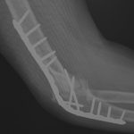
• Skip to main content
  • Skip to primary sidebar
  • Skip to footer
  • General Ortho
  • Procedures
  • Spine
  • Upper Limb
  • Lower Limb
  • Pain
  • Trauma
  • Tumors
  • Newsletter/Updates
  • About Us
  • Contact Us

Bone and Spine

Orthopedic health, conditions and treatment

Hand and Upper Limb

Scapulothoracic Bursitis Causes and Treament

By Dr Arun Pal Singh

scapulothoracic bursitis bursa locations

Scapulothoracic bursitis is inflammation of the scapulothoracic bursae present between the scapula and rib cage. Symptomatic scapulothoracic bursitis is not very common. Another similar but different term used is scapulothoracic crepitus [snapping scapula] and it is important to highlight the difference. Scapulothoracic crepitus is referred to as thumping or popping sounds that occur with scapulothoracic […]

Filed Under: Hand and Upper Limb

Bursae Around Shoulder

By Dr Arun Pal Singh

bursae arund shoulder

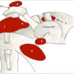
There are 5-6 bursae around the shoulder. Bursae are the synovial fluid-filled sacs that are present between two surfaces that rub on motion. The purpose of the bursa is to reduce friction and protect the moving tissue. Often these are found at tendon-tendon and tendon-bone interfaces. There are five main bursae around shoulder. They include: […]

Filed Under: Hand and Upper Limb

Dorsal Digital Expansion or Extensor Expansion

By Dr Arun Pal Singh

Dorsal Digital Expansion

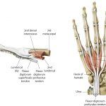
The term dorsal digital expansion refers to the small triangular aponeurosis covering the dorsum of the proximal phalanx with its base at the metacarpophalangeal joint. The main tendon of the extensor digitorum occupies the central part of the expansion and is separated from the metacarpophalangeal joint by a bursa. [Aponeurosis is a flat sheet or […]

Filed Under: Anatomy, Hand and Upper Limb

Anatomy of Muscles of Arm

By Dr Arun Pal Singh

Biceps muscle with both heads of origin

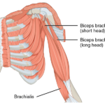
The region between the shoulder joint and the elbow joint is called the upper arm or arm. That between elbow and wrist is called forearm. Brachium and antebrachium are anatomical terms for arm and forearm respectively but are not used that commonly. Muscles of arm can be classified as that of a flexor compartment or […]

Filed Under: Anatomy, Hand and Upper Limb

Rolando Fracture – Presentation and Treatment

By Dr Arun Pal Singh

Rolando Fracture

Rolando fracture is a three-part intraarticular fracture-dislocation of the base of the thumb. This is an unstable injury that requires surgical reduction and fixation. It is named after Silvio Rolando, an Italian surgeon who described it first. Relevant Anatomy, Mechanism of Injury and Pathophysiology The carpometacarpal joint is between the base of thumb and trapezium […]

Filed Under: Hand and Upper Limb

Bennett Fracture Causes, Presentation and Treatment

By Dr Arun Pal Singh

Forces deforming Bennet fracture

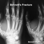
Bennett fracture is an intraarticular fracture involving the base of the thumb and leading to a subluxation of the carpometacarpal joint. It was described initially by Edward Hallaran Bennett in the late nineteenth century. The fracture is inherently unstable and requires adequate reduction and immobilization or fixation. Carpometacarpal joint is critical for pinching and opposition […]

Filed Under: Hand and Upper Limb

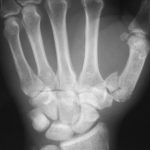
Base of Thumb Fractures and Their Types

By Dr Arun Pal Singh

Rolando Fracture

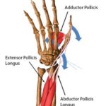
Base of thumb fractures are the extra-articular and intraarticular fractures of base of first metacarpal bone. Thumb is a highly important and efficient digit that makes the human hand highly efficient. An unusable thumb could mean a loss of 40% of hand function. Relevant Anatomy [Read complete hand anatomy] Thumb consists of two phalanges and […]

Filed Under: Hand and Upper Limb

Thumb Injuries Types, Presentation and Treatment

By Dr Arun Pal Singh

Intra-articular Fracture of First Metacarpal Base With Subluxation Of First Metacarpophalangeal Joint

Thumb injuries or fractures and dislocations are one of the common hand injuries. The term thumb injury includes all the injuries from the carpometacarpal joint to the tip of the thumb. There are various types of thumb injuries that come under fractures and dislocations of thumb. Nailbed and fingertip injuries are discussed separately. Thumb is […]

Filed Under: Hand and Upper Limb

Skier’s thumb – Causes, Symptoms and Teatment

By Dr Arun Pal Singh

skier's thumb or gamekeeper's thumb

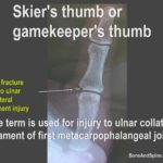
Terms Skier’s thumb and Gamekeeper’s thumb are used for injuries to the ulnar collateral ligament of first metacarpal joint. While Gamekeeper’s thumb was the older term, Skier’s thumb is used more commonly. Ulnar collateral ligament injury of the metacarpal joint was found earlier in people of Europe who wrung the neck of the game like […]

Filed Under: Hand and Upper Limb

Jersey Finger – Causes, Symptoms and Treatment

By Dr Arun Pal Singh

Jersey finger

Jersey finger refers to the avulsion of the flexor digitorum profundus tendon off of its insertion at the volar aspect of the distal phalanx due to forced hyperextension of the distal interphalangeal joint. It is a type of zone I tendon injury. It is also called a sweater finger or rugby finger and typically occurs […]

Filed Under: Hand and Upper Limb

  • Go to page 1
  • Go to page 2
  • Go to page 3
  • Interim pages omitted …
  • Go to page 12
  • Go to Next Page »

Primary Sidebar

Lumbar plexus details, PD wiki

Lumbosacral Plexus Anatomy

Lumbosacral plexus is formed anterior or ventral rami of L1–S3 roots. All major nerves of the lower extremity are derived from lumbosacral plexus. Though functionally considered as one entity, the lumbosacral plexus is usually thought of anatomically as an upper lumbar plexus and a sacral plexus also called lower lumbosacral plexus [also receives lumbar contribution]. […]

Xray of Congenital Radioulnar Synostosis Showing Fused Ulna and Radius

Congenital Radioulnar Synostosis Presentation and Treatment

In congenital radioulnar synostosis, there is congenital fusion of the proximal ends of the radius and ulna [a bony bridge between the proximal radius and ulna]. This results in the fixation of the forearm in varying degrees of pronation. That means the upper end of both the bones, radius, and ulna are fused, making it […]

Slipped Cpper Femoral Epiphysis

Slipped Femoral Capital Epiphysis

Slipped femoral capital epiphysis is the disorder of the proximal femoral physis that leads to slippage of the epiphysis relative to the femoral neck. It is one of commonest hip abnormalities in adolescence. It affects both the hips in about 20% of cases. The incidence for slipped capital femoral epiphysis is about 10 cases per […]

External Fixator Applied To Tibia

External Fixators – Concept and Use

An external fixator is a useful tool in the management of fractures and certain difficult orthopedic problems such as limb length discrepancy. External fixation a useful tool in fracture management and in the case of pelvic fracture it may be a primary life-saving device With external fixation, pins and/or wires are percutaneously inserted into the […]

Periprosthetic Fracture Femur in a patient who had undergone bipolar hemiarthroplasy previously for fracture of neck of femur

Hip Injuries Xrays and Photographs

Hip injuries consist of injuries to the acetabulum, lower pelvis, intertrochanteric fractures, fracture of neck of femur, fracture of the femoral head, subtrochanteric fractures and hip dislocations. A collection of x-rays and clinical photographs of hip injuries is being presented. Image 1 –  Xray of Oblique Subtrochanteric Fracture of Femur With Dynamic Hip Screw and […]

Radius fracture Fracture fixed with DCP

Fracture Reduction and Fixation – Principles, Types and Application

Fracture reduction treatment has evolved from its earlier days and now includes many modalities.  This article describes the basic terms of reduction and fixation. Closed Reduction Closed reduction of the fracture means reducing the fracture fragments into acceptable anatomical position.  After reduction has been achieved by manipulation and maneuvers, the alignment is generally maintained by […]

scapulothoracic bursitis bursa locations

Scapulothoracic Bursitis Causes and Treament

Scapulothoracic bursitis is inflammation of the scapulothoracic bursae present between the scapula and rib cage. Symptomatic scapulothoracic bursitis is not very common. Another similar but different term used is scapulothoracic crepitus [snapping scapula] and it is important to highlight the difference. Scapulothoracic crepitus is referred to as thumping or popping sounds that occur with scapulothoracic […]

Browse Articles

Footer

Pages

  • About
    • Policies
    • Contact Us

Featured Article

Shoulder Dislocation Presentation and Treatment

Shoulder dislocation refers to dislocation of the glenohumeral joint. Shoulder dislocation is a common injury and occurs more in young adults.  … [Read More...] about Shoulder Dislocation Presentation and Treatment

Search Articles

© Copyright: BoneAndSpine.com