Last Updated on October 28, 2023
Lumbosacral plexus is formed anterior or ventral rami of L1–S3 roots. All major nerves of the lower extremity are derived from lumbosacral plexus.
Though functionally considered as one entity, the lumbosacral plexus is usually thought of anatomically as an upper lumbar plexus and a sacral plexus also called lower lumbosacral plexus [also receives lumbar contribution]. Some authors also call the sacral plexus as simply lumbosacral plexus and such usage can cause confusion. It is better to refer to them as lumbar and sacral plexus.
The pudendal plexus is a term used for a compound structure consisting of sacral spinal nerves. It gets a contribution from the S2-S4 segment and is also called S2-S4 of the lumbosacral plexus. It is part of lumbosacral plexus.
Anatomy of Lumbar Plexus
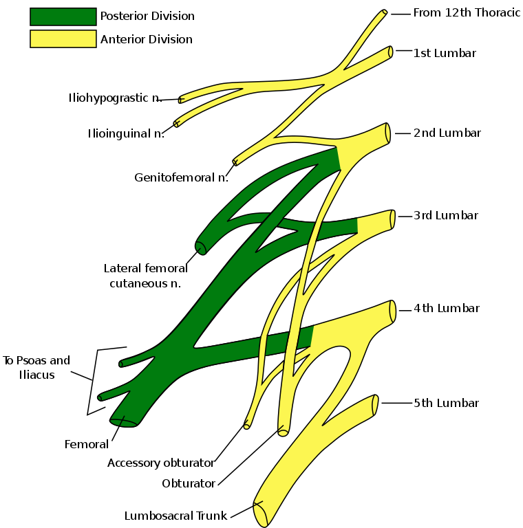
The lumbar plexus is formed by the divisions of the ventral rami of L1-L4 nerves. It also gets contributions from subcostal nerve which is last thoracic nerve [T12].
The ventral rami of the fourth lumbar nerve pass communicating branches, the lumbosacral trunk, to the sacral plexus.
The plexus is formed lateral to the intervertebral foramina and passes through psoas major.
It directly gives motor branches are distributed directly to psoas major. Larger branches leave the muscle at various sites to run obliquely down through the pelvis and pass to thigh under the inguinal ligament. The exception is the obturator nerve which leaves the pelvis through the obturator foramen.
Branches of Lumbar Plexus
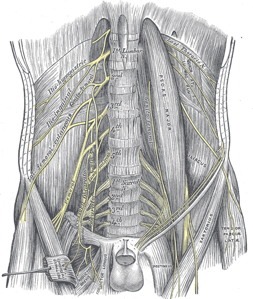
Iliohypogastric and Ilioinguinal Nerves
These two paired nerves, derived from the L1 root
Iliohypogastric nerve contains fibers from T12 also] runs posterior to the psoas major on its proximal lateral border to run laterally and obliquely on the anterior side of quadratus lumborum muscle. Lateral to this muscle, these nerves pierce the transversus abdominis to run above the iliac crest between that muscle and abdominal internal oblique. It supplies muscular innervation to the transverse and internal oblique muscles.
The iliohypogastric nerve also gives a sensory branch to the skin of the lateral hip. Its terminal branch then runs parallel to the inguinal ligament to exit the aponeurosis of the abdominal external oblique above the external inguinal ring where it supplies the skin above the inguinal ligament (the hypogastric region) with the anterior cutaneous branch.
The ilioinguinal nerve closely follows the iliohypogastric nerve on the quadratus lumborum, but then passes below it to run at the level of the iliac crest. With similar course, it also supplies motor branches to both transversus abdominis and internal oblique muscles.
Ilioinguinal nerve supplies sensation to
- An area of skin over the inguinal ligament
- A small area of skin over the rostral medial thigh
- The upper part of the scrotum in males or labia in females.
Genitofemoral Nerve [L1,L2]
This small nerve is derived from both the L1 and L2 fibers. The genitofemoral nerve pierces psoas major anteriorly below the former two nerves to immediately split into two branches that run downward on the anterior side of the muscle. [ a genital and a femoral branch] at the level of the medial inguinal ligament.
The genital branch differs in males and females. In males, it runs in the spermatic cord and in females in the inguinal canal together with the teres uteri ligament. The genital branch provides muscular innervation to the cremasteric muscles in males and sensation to the skin over the lower part of the scrotum in males or labia in females.
Lateral Cutaneous Nerve of Thigh or Lateral Femoral Cutaneous Nerve [L2–L3]
The lateral femoral cutaneous nerve is a pure sensory nerve that is derived from the L2–L3 roots and emerges laterally from the psoas muscle, and then crosses obliquely toward the anterior superior iliac spine (ASIS) where it passes under the inguinal ligament.
It is here at the ASIS and inguinal ligament that the nerve is susceptible to injury and compression. [Meralgia Paresthetica].
The nerve emerges from the underlying fascia about 10–12 cm distally and divides into anterior and posterior branches that supply sensation to a large oval area of skin over the lateral and anterior thigh.
Obturator Nerve [ L2, L3, L4]
Obturator nerve is formed by the anterior divisions of the L2–L3–L4 anterior rami. The obturator nerve leaves the lumbar plexus and descends behind psoas major on it medial side and leaves the pelvic area through the obturator canal.
After giving a motor branch to obturator externus in the thigh, it divides into an anterior and a posterior branch. The continue distally, separated by adductor brevis. Obturator nerve supplies thigh adductors namely adductor longus, adductor brevis, adductor magnus, adductor minimus, and gracilis. It also gives a motor branch to pectineus.
A sensory branch from the anterior division passes along the anterior border of gracilis and supplies the skin on the medial distal part of the thigh.
Femoral Nerve [L2, L3, L4]
The femoral nerve is the largest and longest of the lumbar plexus nerves..
The three posterior divisions of anterior rami of the L2–L3–L4 roots unite to form the femoral nerve, which runs through the pelvis in a groove between psoas major and iliacus. It gives off branches to both muscles, and exits the pelvis through and enters into the thigh under the inguinal ligament.
It divides into sensory and muscular branches and the saphenous nerve which is a sensory terminal branch that supplies calf and hindfoot.
It provides motor supply to the iliopsoas, pectineus, sartorius, and quadriceps muscles and sensory innervation to the anterior thigh, posterior lower leg, and hindfoot.
Anatomy of Sacral Plexus or Lower Lumbosacral Plexus
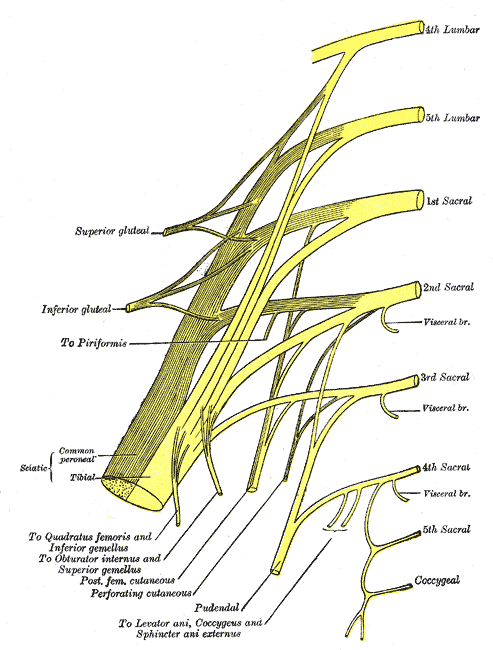
The sacral plexus (plexus sacralis) is s part of the larger lumbosacral plexus. The sacral plexus is derived from the anterior rami of spinal nerves L4, L5, S1, S2, S3, and S4.
The origin may be shifted upward [prefixed] or downward [postfixed] to the extent of one spinal nerve. Following structures participate in the formation of lumbosacral plexus.
- Lumbosacral trunk
- Anterior division of the first sacral nerve
- Portions of the anterior divisions of the second and third sacral nerves
Lumbosacral trunk is the connection between the lumbar and sacral plexus.
It is formed by the whole of the anterior division of the L5 and a part of that of the L4 nerve.
The lumbosacral trunk appears at the medial margin of the psoas major and runs downward over the pelvic brim to join the first sacral nerve.
Sacral contributing rami receive a gray ramus communicans from the corresponding ganglion of the sympathetic trunk.
Also S2,3,4 give a pelvic splanchnic nerve is given to the inferior hypogastric plexus providing parasympathetic fibers to supply the hindgut and the pelvic viscera.
The sacral plexus lies in the back of the pelvis between the piriformis muscle and the pelvic fascia.

Anterior to the plexus are the internal iliac artery, internal iliac vein, the ureter, and the rectum.
The superior gluteal artery and vein usually run between the lumbosacral trunk and the first sacral nerve
The inferior gluteal artery and vein often run between the second and third sacral nerves.
All the nerve roots entering the plexus split into anterior and posterior divisions, and converge toward the lower part of the greater sciatic foramen, and unite to form a flattened band.
The branches of sacral plexus arise from this band and the band itself is continued as the sciatic nerve.
The sciatic nerve splits on the back of the thigh into the tibial nerve and common fibular nerve. Sometimes, these two nerves arise separately from the plexus.
Nerves from Sacral Plexus
Nerve to the Quadratus Femoris and Gemellus Inferior [L4-S1]
This nerve arises from the anterior divisions of L4, L5, and S1 nerve roots. It leaves the pelvis through the greater sciatic foramen, below the piriformis, and runs down in front of the sciatic nerve, the gemelli, and the tendon of the obturator internus.
It then enters the anterior surfaces of the quadratus femoris and gemellus inferior muscles.
It also gives an articular branch to the hip joint.
Nerve to the Obturator Internus and Gemellus Superior [L5-S2]
This arises from the anterior division of the L5, S1 and S2 roots.
It leaves the pelvis through the greater sciatic foramen below the piriformis. It enters the upper part gemellus superior muscle and gives off the branch. It enters the perineum through the lesser sciatic foramen and supplies the pelvic surface of the obturator internus muscle.
Nerve to the Piriformis [S1, S2]
It arises from S1 and S2 nerve and enters the second sacral nerve roots and enters the anterior surface of the muscle.
Superior Gluteal Nerve [L4-S] and Inferior Gluteal Nerves [L5-S2].
The superior gluteal nerve is derived from L4–L5–S1 fibers, leaves the greater sciatic foramen above the piriformis, accompanied by the superior gluteal vessels, and divides into a superior and an inferior branch.
It supplies the tensor fascia latae, gluteus medius, and gluteus minimus muscles but carries no sensory fibers for skin.
The superior branch is accompanied by the upper branch of the deep division of the superior gluteal artery and ends in the gluteus minimus.
The inferior division gives filaments to the gluteus medius and minimus and ends in the tensor fascia lata.
.
The inferior gluteal nerve, derived from L5–S1–S2 fibers, supplies only the gluteus maximus muscle, which subserves extension of the hip joint.
It leaves the pelvis through the greater sciatic foramen, below the piriformis.
It branches to enter the deep surface of the gluteus maximus.
Posterior Femoral Cutaneous Nerve [S1-S3]
It arises partly from the posterior divisions of the first and second sacral nerve roots and from the anterior divisions of the second and third sacral nerve roots. S2 is the main contributor but also has a component from S1 and S3.
It leaves the pelvis adjacent to the sciatic nerve to supply sensation to the skin of the perineum and posterior surface of the lower portion of the buttock, thigh, and leg
Sciatic Nerve Tibial part: [L4-S3], Common Fibular part [L4-S2]
The sciatic nerve, which receives innervation from the L4–S3 roots. It is the largest nerve in the body, measuring 2 cm in breadth, and is also the continuation of the flattened back of the sacral plexus. The sciatic nerve passes out of the pelvis through the greater sciatic foramen, below the piriformis muscle.
It descends between the greater trochanter of the femur and the tuberosity of the ischium and along the back of the thigh to about its lower third, where it divides into tibial and common fibular (peroneal) nerves.
However, the division may take place at any point between the sacral plexus and the lower third of the thigh.
The sciatic nerve supplies the hamstring muscles and lateral division of adductor magnus muscle.
Through its branches tibial nerves and common fibular nerves it also supplies skin and muscles of leg and foot.
Clinical Significance of Lumbosacral Plexus
Lumbosacral plexus may be involved in many disorders, commonly called as lumbosacral plexopathy. It may be injured during birth, trauma or in chronic diseases like diabetes.
For correct diagnosis, it is important to differentiate between nerve disorders and plexus disorders.