• Skip to main content
  • Skip to primary sidebar
  • Skip to footer
  • General Ortho
  • Procedures
  • Spine
  • Upper Limb
  • Lower Limb
  • Pain
  • Trauma
  • Tumors
  • Newsletter/Updates

Bone and Spine

Orthopedic health, conditions and treatment

Patellar Translation or TT TG Distance

By Dr Arun Pal Singh

In this article
    • Method of Measurement of TT-TG distance
    • Inference of Patellar Translation

Patellar translation is the distance between tibial tubercle and trochlear groove and is also called TT-TG distance. It is measured on CT or MRI by overlapping or superimposing axial images of the femoral condyles and tibial tuberosity.

The measurements are used to quantify patellar instability.

The position of the tibial tubercle is crucial for the inferolateral force vector of the patella [a force that acts on inferior and lateral direction].

In a normal joint, the tibial tuberosity lies vertically under the femoral sulcus, directing the force vector inferiorly during knee bending.

But if there is if there is excessive lateralization of the tibial tuberosity, the patella is pulled laterally during flexion as the tuberosity is lateral and thus pull would also be.

If this is the cause of patellar instability, realignment of tibial tuberosity may help the patient with patellar instability.

Method of Measurement of TT-TG distance

Calculation of Tibial Tuberosity - Trochlear Groove Distance or Patellar Translation
Calculation of Tibial Tuberosity – Trochlear Groove Distance, Image Credit: RSNA

?For calculation of patellar translation or TT TG distance, axial images of tibia with tibial tuberosity are taken. Axial images of femoral condyle are also taken.

Then axial images of femoral condyles and tibial tuberosity are superimposed.

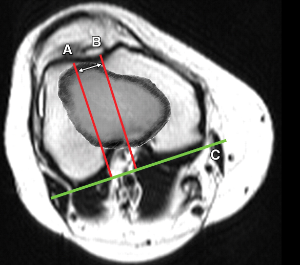
A line is drawn along the posterior femoral condyles.[line C]

Then two lines are drawn perpendicular to this line. [A and B]

The first line bisects the tibial tuberosity[TT] and the second  bisecting the trochlear groove sulcus [TG]

The distance between TT and TG line is  TT-TG distance or patellar translation. In the image distance between A and B.

Inference of Patellar Translation

A distance of <15 mm is considered normal,

15-20 mm is considered borderline

a distance >20 mm is considered abnormal. A tibial tubercle–trochlear groove distance of more than 20 mm is nearly always associated with patellar instability

The distance has been shown to be slightly less when measured on MRI than CT.

The measurement of the lateral distance between the tibial tubercle and the trochlear groove is less accurate in individuals with severe trochlear dysplasia as the deepest point of the trochlea cannot be defined.


Spread the Knowledge
  • 8
    Shares
  •  
    8
    Shares
  • 7
  •  
  • 1
  •  

Filed Under: Knee

ebook on fracture care

About Dr Arun Pal Singh

Arun Pal Singh is an orthopedic and trauma surgeon, founder and chief editor of this website. He works in Kanwar Bone and Spine Clinic, Dasuya, Hoshiarpur, Punjab.

This website is an effort to educate and support people and medical personnel on orthopedic issues and musculoskeletal health.

You can follow him on Facebook, Linkedin and Twitter

Reader Interactions

Leave a Reply Cancel reply

Your email address will not be published. Required fields are marked *

This site uses Akismet to reduce spam. Learn how your comment data is processed.

Primary Sidebar

Accessory bone Os velanium of the foot

Os Vesalianum – The Accessory Bone of Fifth Metatarsal

What is Os Vesalianum Os vesalianum pedis is an accessory bone of the foot located proximal to the fifth metatarsal and found within the peroneus brevis tendon. It is a rare occurrence. The reported incidence is between 0.1% and 0.4%. This ossicle is without any symptoms generally but often can become painful. It is often […]

Dystrophic nail in nail patella syndrome

Nail Patella Syndrome

Nail Patella syndrome is an autosomal dominant condition characterized abnormalities of the nails, knees, elbows, and pelvis. The features vary in severity from individual to individual, even among members of the same family. The classical description is of a tetrad of nail dysplasia, patellar aplasia-hypoplasia, elbow joint dysplasia, and iliac horns. Nail abnormalities are seen […]

Ulnar Claw hand

Ulnar Neuropathy [Compressive] – Causes and Treatment

Ulnar neuropathy refers to compression of the ulnar nerve, a continuation of the medial cord of the brachial plexus and a mixed nerve that supplies muscles of the forearm, and skin over medial part of hand and digits. Ulnar neuropathy is the second most common entrapment neuropathy of the upper limb. There are various sites […]

genu varum rickets

Genu Varum or Bow Legs Causes and Treatment

This gives the limb an overall the appearance of an archer’s bow. This condition may present from infancy through adulthood and has a variety of causes. The problem may be unilateral which would result in functional leg length discrepancy or bilateral. It is interesting to note that almost all babies are born bow-legged. By the time […]

Lateral view of ankle joint

Ankle Joint Anatomy

The ankle joint is a hinged synovial joint. In an absolute sense, its primary movements are moving the foot up and down, called dorsiflexion and plantar flexion respectively. However, this joint works in tandem with subtalar joints [talocalcaneal and talocalcaneonavicular], thus adding to the range of motion of this ankle-subtalar joint complex. Anatomically subtalar (talocalcaneal) […]

Finkelstein test for dequervain tenosynovitis

De Quervain Tenosynovitis Symptoms and Treatment

De Quervain tenosynovitis is a tenosynovitis of the tendons within the first extensor compartment at the wrist resulting in pain during thumb motion. Other names fo De Quervain tenosynovitis are De Quervain syndrome, BlackBerry thumb, gamer’s thumb, washerwoman’s sprain, radial styloid tenosynovitis, de Quervain disease, de Quervain’s stenosing tenosynovitis, mother’s wrist, or mommy’s thumb. [1] […]

Monteggia Fracture Dislocation

Monteggia Fracture Causes, Types, Presentation and Treatment

Monteggia fracture-dislocations consist of a fracture of the proximal 1/3 of the ulnar shaft with concomitant dislocation/instability of the radial head. Often the ulnar fracture is obvious but radial head dislocation may be subtle and prone to be overlooked. Therefore high injury of suspicion is required for correct diagnosis and it is important to view […]

Browse Articles

Footer

Pages

  • About
    • Privacy Policy
    • Disclaimer
    • Contact

Featured Article

Anatomy of Ulna Bone

Ulna is a long bone in the forearm. It lies medially and parallels to the radius, the other bone in the forearmIt acts as stabilizing the bone, … [Read More...] about Anatomy of Ulna Bone

Search Articles

© Copyright: BoneAndSpine.com