• Skip to primary navigation
  • Skip to main content
  • Skip to primary sidebar
  • Home
  • TeleConsult
  • About
  • Newsletter/Updates
  • Contact Us
  • Policies

Bone and Spine

Orthopedic health, conditions and treatment

  • General Ortho
  • Procedures
  • Spine
  • Upper Limb
  • Lower Limb
  • Pain
  • Trauma
  • Tumors

Rolando Fracture – Presentation and Treatment

By Dr Arun Pal Singh

In this article
    • Relevant Anatomy, Mechanism of Injury and Pathophysiology
    • Clinical Presentation
    • Imaging
    • Differential Diagnosis
    • Treatment
    • Complications
    • References
      • Related

Rolando fracture is a three-part intraarticular fracture-dislocation of the base of the thumb. This is an unstable injury that requires surgical reduction and fixation. It is named after Silvio Rolando, an Italian surgeon who described it first.

Relevant Anatomy, Mechanism of Injury and Pathophysiology

The carpometacarpal joint is between the base of thumb and trapezium bone.

Carpal Bones or Bones of the carpus - wrist
Carpal Bones

Muscles of thumb act as deforming forces to base of thumb fractures

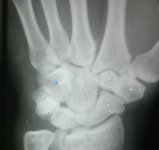
The joint allows motion parallel and perpendicular to the plane of the palm. Thumb function constitutes about 50% of hand function as a whole. The thumb metacarpal base is a unique that it allows a wide range of motion while maintaining stability for grasp and pinch.

Therefore, it  is very important that accurate reduction and aggressive rehabilitation is carried when

Base of thumb fractures are mainly extraarticular and intraarticular. Intraarticular fractures are namely Bennet fracture, Rolando fracture, and comminuted fractures.

The Rolando fracture usually occurs due to axial loading in a partially flexed metacarpal, such as a fistfight. It can also occur as a result of indirect forces such as falling on a thumb.

Punching is the most common mechanism but other causes like motor vehicle accidents, work-related injuries and falls are also common.

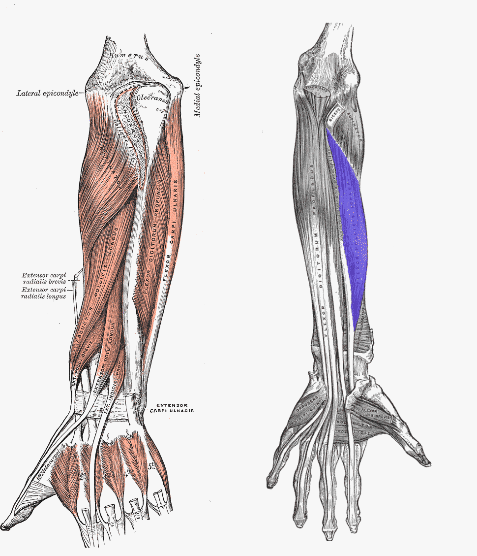
The resting tone present in the multiple tendons that act on the thumb further lead to the displacement.

These include extensor pollicis brevis and extensor pollicis longus which shorten the thumb ray along with pull from the flexor pollicis longus.

Extensor muscles to the thumb

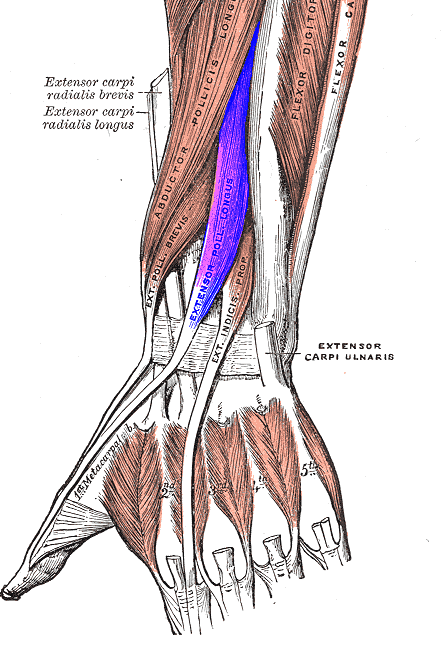
The adductor pollicis pulls the distal metacarpal toward the palm and in conjunction with the abductor pollicis longus [acts on the metacarpal base] leads to varus at the metaphyseal-diaphyseal junction.

The fracture line is typically T- or Y-shaped.

Clinical Presentation

After the injury, the patient presents with swelling and pain in the base of the thumb region. The region is tender and movements are painful.

Neurovascular and tendon injuries are not commonly associated with this fracture.

Imaging

The fracture is confirmed by x-ray imaging.

Anteroposterior (AP) and lateral radiographs are the routine x-rays sought.  If the routine x-rays are not able to reveal the full extent of comminution, a Robert radiograph (a hyperpronated view of the thumb base) can be tried.

Rolando fracture is very similar to Bennett fracture but it divides the metacarpal base into two, three and sometimes many fragments. Rolando fracture is also called as Y or T shape fracture.

Rolando Fracture
Rolando Fracture, Image from Wikipedia, GNU license

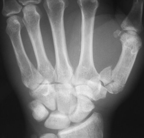
CT is able to display the fracture configuration better.

Differential Diagnosis

  • Bennett Fracture
  • Extra-articular fractures through the first metacarpal base
  • Comminuted fractures of -part intra-articular fracture through the first metacarpal base

Treatment

Most of the Rolando fractures are displaced and therefore treated by surgery. Only minimally displaced fractures should be considered for thumb spica.

All fractures with articular stepoff more than 1-2 mm would require surgical treatment.

Various treatment options can be used as follows

  • Large articular fragments – plate-and-screw fixation.
  • Massive comminution-  traction or ligamentotaxis and external fixators.
  • Open fractures – Prefer pin fixation or external fixation

After the surgery and sufficient union, the patient is put on exercises which include stretching and strengthening exercises of wrist, hand and thumb in particular. These include

  • Opposition Stretch:
  • Wrist Flexion-extension
  • Grip Strength.
  • Finger Stretch

Complications

Complications are often directly related nature of the injury and the extent of the comminution. Usually, these are

  • Pin-tract infection
  • Screw pullout and loss of fixation
  • Neural damage
  • Joint stiffness

References

  1. Breen TF, Gelberman RH, Jupiter JB. Intra-articular fractures of the basilar joint of the thumb. Hand Clin. 1988 Aug. 4 (3):491-501
  2. Mumtaz MU, Ahmad F, Kawoosa AA, Hussain I, Wani I. Treatment of Rolando Fractures by Open Reduction and Internal Fixation using Mini T-Plate and Screws. J Hand Microsurg. 2016 Aug. 8 (2):80-5.
  3. Gelberman RH, Vance RM, Zakaib GS. Fractures at the base of the thumb: treatment with oblique traction. J Bone Joint Surg Am. 1979 Mar. 61 (2):260-2

Related

Spread the Knowledge


Filed Under: Hand and Upper Limb

About Dr Arun Pal Singh

Arun Pal Singh is an orthopedic and trauma surgeon, founder and chief editor of this website. He works in Kanwar Bone and Spine Clinic, Dasuya, Hoshiarpur, Punjab.

This website is an effort to educate and support people and medical personnel on orthopedic issues and musculoskeletal health.

You can follow him on Facebook, Linkedin and Twitter

Primary Sidebar

Browse Articles

Shin Splints are Common in Runners

Shin Splints or Medial tibial stress syndrome

Shin splints or Medial tibial stress syndrome is an overuse injury or repetitive stress injury of the shin area [tibial bone]. These affect persons who engage in running and jumping. It affects about 15% of all running-related injuries. It is most often found in runners, and persons playing other sports like football, basketball, soccer, and […]

Foot Xray Showing Marked Bones and Foot Regions

Normal Foot X-ray – Findings and Discussion

Normal Foot Xray Xrays are basic investigations for orthopedic problems. To find the abnormal one must have an idea of normal. This is how a normal foot x-ray looks like. The present image shows an anteroposterior and oblique view x-ray of the left foot. On the left side of the plate, there is an oblique […]

clavicle bone or collarbone as seen on surface

Clavicle Anatomy and Attachments

The clavicle elongated, S-shaped bone that lies horizontally between the sternum and the acromial end of the scapula. It is an important connection between the axial skeleton and the pectoral girdle. The pectoral girdle or the shoulder girdle is the term used for bones in the appendicular skeleton which connects to the arm on each […]

Different ratios for calculating patellar height

Patella Baja or Low Lying Patella

Patella Baja or patella infera is an abnormally low lying patella [compare with patella alta, the high riding patella]. It is associated with a restricted range of motion, crepitations, and retropatellar pain. Patella baja could be congenital, acquired, or a combination of the two is commonly encountered in total knee arthroplasty. Any cause that would lead to […]

wound-dressings

Wound Healing Process and Types

Wound healing is a complex process taken up by the body to repair the defect by restoring cellular structure and tissue layers. Wound healing process has distinct phases which are Hemostasis Inflammatory Phase Proliferative or Granulation Phase Remodeling Phase Hemostasis Some authors consider this a separate entity and do not include in phases of wound […]

purine rich foods

Purines and Foods Rich in Purines

Purines are natural substances that form part of the chemical structure of our genes and the genes of plants and animals. Though these are present in many foods, a relatively small number of foods contain concentrated amounts of them. By and large, these foods are also high-protein foods. Examples are meats like kidney, fish like […]

Fresh and Healed Blisters on Leg

Complications of Plaster Cast

A plaster cast is given for the treatment of fractures and other orthopedic ailments. Though a very safe mode of treatment, complications of plaster cast may occur. The main complications of plaster cast are stiff joints, muscle wasting, and impaired circulation. Physiotherapy and good nursing can help reduce these complications and speed the final recovery. […]

© Copyright: BoneAndSpine.com
Manage Cookie Consent
The site uses cookies. Please accept cookies for a better visiting experience.
Functional Always active
The technical storage or access is strictly necessary for the legitimate purpose of enabling the use of a specific service explicitly requested by the subscriber or user, or for the sole purpose of carrying out the transmission of a communication over an electronic communications network.
Preferences
The technical storage or access is necessary for the legitimate purpose of storing preferences that are not requested by the subscriber or user.
Statistics
The technical storage or access that is used exclusively for statistical purposes. The technical storage or access that is used exclusively for anonymous statistical purposes. Without a subpoena, voluntary compliance on the part of your Internet Service Provider, or additional records from a third party, information stored or retrieved for this purpose alone cannot usually be used to identify you.
Marketing
The technical storage or access is required to create user profiles to send advertising, or to track the user on a website or across several websites for similar marketing purposes.
Manage options Manage services Manage vendors Read more about these purposes
View preferences
{title} {title} {title}
 

Loading Comments...