Last Updated on November 22, 2023
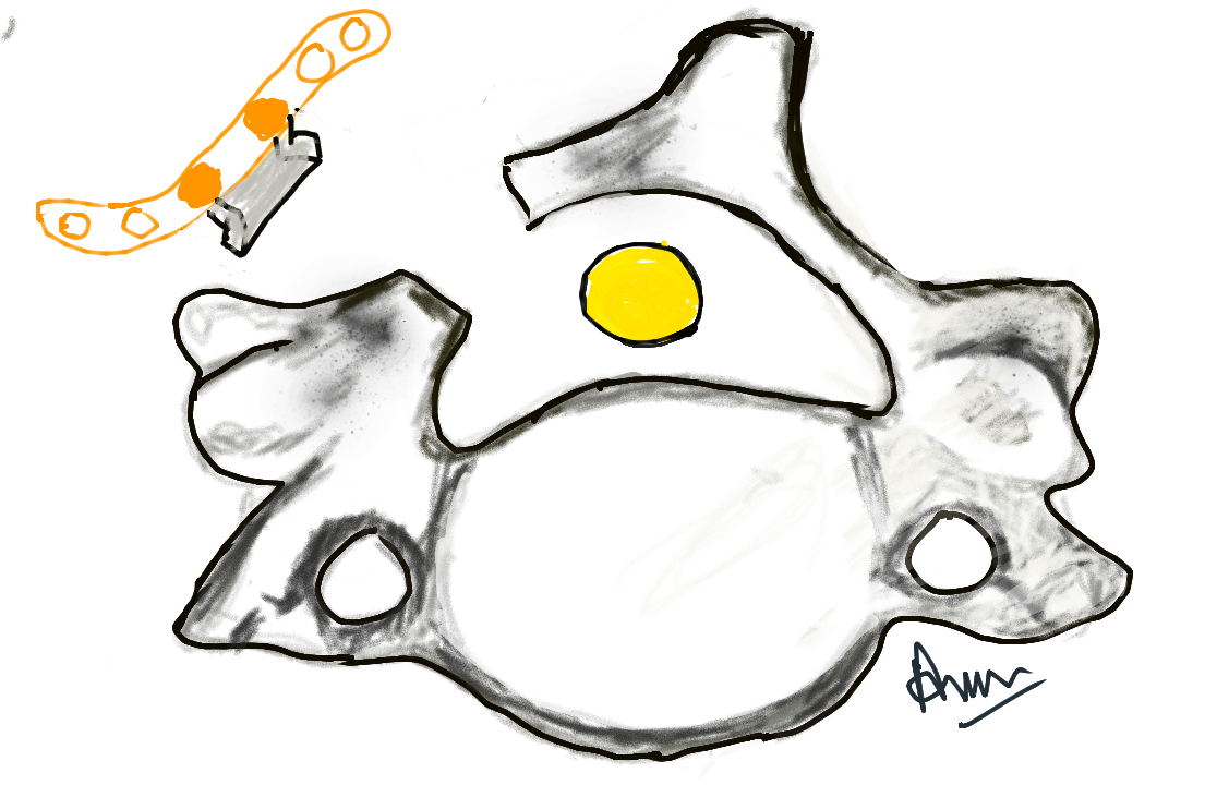
Laminoplasty is a process of increasing the space available for the spinal cord by reconstruction of the laminar arch via a posterior approach.
It is done by hinging or opening the lamina to allow access to the canal and may include augmentation or reconstruction of the spinal canal.
Laminectomy is just removal of the lamina and it also the medial posterior tension band structures such as the supraspinous ligament, interspinous ligament, ligamentum flavum, spinous processes, and the muscle insertions on the midline.
But the laminectomy is associated with a risk of postoperative instability and kyphosis. The risk of postlaminectomy instability is highest in the cervical spine and lowest in the lumbar spine
A laminoplasty, on the other hand, is able to create more space for the spinal cord and nerve roots and able to preserve the stability of the spine while preserving the motion [no fusion required]
Cervical laminoplasty had been developed in Japan for many years to overcome the drawbacks of laminectomy for the treatment of ossification of the posterior longitudinal ligament (OPLL) and degenerative spinal canal stenosis.
Laminoplasty on the thoracic and lumbar spine was first described by Raimondi and colleagues in 1976.
It has been used commonly for access to the spinal cord for indications including syringomyelia, tumors, functional, and painful surgery.
Indications for Laminoplasty
- Cervical Spine
- Cervical myelopathy due to developmental canal stenosis [AP diameter less than 12mm]
- Spondylotic changes at more than three segments in cervical spine
- Ossification of the posterior longitudinal ligament compressing the spinal cord at multiple levels.
- The thoracic and lumbar spine
- To access underlying spinal disease (eg, tumor, syringomyelia).
Prerequisite and Contraindications
Prerequisites
- A physiological spinal alignment the prerequisite for a laminoplasty.
- No instability
- Positive K- line
- A virtual connecting the midpoints of the anteroposterior diameter of the spinal canal at C2 and C7 in a plain lateral radiogram.
- Positive – The peak of OPLL does not exceed the K-line
- Negative – Tthe peak of OPLL exceeds the K-line
- Laminoplasty should not be used if the K-line is negative [Rather Decompression and fusion with instrumentation]
Contraindications
- Spinal pathologies, which are best treated by an anterior approach
-
- Cervical disc disease
- Traumatic vertebral body fracture with canal compression)
- Cervical spondylolisthesis
- Spinal instability of the affected segment
- Kyphotic deformity
- Patients medically unfit for surgery
- Cervical kyphotic deformity
Patient Assessment and Planning
Surgical considerations should include-
- Assessment of bone quality imaging
- Spinal curvature and space available for cord
- Assessment of degenerative changes and K-line
- Instability [hypermobility] using flexion-extension radiographs
- Assessment of foraminal stenosis [to assess the need for foraminotomy]
A surgical microscope should be used to improve visualization.
The anesthetist should assess the need of awake-fiberoptic intubation to prevent neurological injury [due to hyperextension of the neck needed for routine intubation].
Consider the use of somatosensory-evoked potentials and motor-evoked potentials and accordingly use anesthetic agents [prefer intravenous over inhalation]
The patients are operated in a prone position.
Cervical Laminoplasty Procedure
Usually, the levels involved in laminoplasty are C3-C7. C2 involvement can lead to greater instability and should be considered on an individual basis.
- Midline incision
- Midline incision to the nuchal ligament to preserve its muscle insertions ( trapezius, rhomboid minor, splenius capitis, and serratus posterior superior).
- Expose the spinous processes, leave the supraspinous ligament intact
- Perform a subperiosteal dissection down to the laminae bilaterally while lateralizing the paraspinal muscles.
- Stay on the lateral aspect of the spinous process
- Perform dissection only along with the bony resistance.
- Visualization of the medial half of the facet joints marks the lateral most limit of the dissection
- Retract using spinal retractors
- Identify the inferior border of the lowest lamina is identified using a Penfield dissector.
- Remove a few millimeters of bone on each side with a Kerrison punch to create an entry point.
- To preserve the alignment, a temporary longitudinal miniplate is applied as medially as possible.
- The laminar flap can be removed once troughs are cut.
- Using a drill or craniotome draw through along a line just medial to the facet joints and parallel to the spinous processes to create a laminar trough. A rose burr may be used in case the craniotome demands too much force
- On reaching the inner layer, complete the cutting with a diamond burr.
- Once the troughs are cut, the laminar flap can be removed. The ligamentum flavum is not removed yet and left in place for protection during bone work
- Copious irrigation is done throughout.
Different Types of Cervical Laminoplasty and Their Procedural Variations
There is procedural variation depending on the type of laminoplasty being done. These are mentioned briefly below.
Z-Laminoplasty
The spinous processes are removed and the laminae thinned using a diamond burr. The laminae are split in a transverse direction and reapproximated in an augmented fashion using nonresorbable sutures.
Hinged-door Laminoplasty
One side of the troughs is opened and the laminae are hinged to other side. The hinge is made on the side where the lesser need for decompression is estimated i.e. opposite to the side of the main symptoms.
Holes are drilled through the lateral mass of the hinge-side. Using a non-resorbable suture, lateral retraction of the hinged laminae is maintained.
An interposition graft (rib, spinous process) can be used as a spacer between the medial part of the lateral mass and the laminae.
Following videos gives an overview of the procedures
Midline-enlargement laminoplasty
Both lateral troughs are drilled without penetrating the inner cortical layer of the laminae. Lamina is split along the midline by dividing the spinous process with a drill or saw.
Both the drilled troughs are used as hinges.
Once the spinous process is split, the hemi-arches are reflected laterally to each side. A ceramic, hydroxyapatite or bone graft is wedged into place and secured to place to keep both laminar wings apart.
Bilateral miniplate laminoplasty
It is also based on opening the bilateral laminar troughs and the arches are removed for reconstruction with osteosynthetic mini plates.
The laminotomy block can be removed to allow access to the spinal canal for further decompression.
The anterior border of the laminar arch is trimmed and the flap is secured to the lateral masses using mini plates.
Proximal [cephalad] or distal [caudal] ends of the flap are sutured to the supraspinous ligaments on either end.
Another variation of the procedure involves reflecting the laminotomy block can be reflected upward when both troughs are cut by letting the flap remained attached to the superior supraspinatus ligament.
In all procedures, after surgery, 6-week immobilization using a rigid collar or brace fis given for proper healing.
Thoracolumbar laminotomy
- Midline incision till the thoracolumbar fascia
- Subperiosteal dissection down to the laminae
- The costotransverse process indicates the position of a pedicle.
- Remove a few millimeters of bone between facet joint laterally and spinous process medially.
- Leave the ligamentum flavum for protection until the bone work is done.
- Link the spinous processes using titanium mesh plates prior to laminotomy
- Preserves the length and angulation of the spinous processes.
- Plates removed after laminae have been fixed back into place.
- Divide the ligamentum interspinosum of the most caudal vertebra is divided sharply.
- Keep the cranialmost interspinous ligament attached if the flap is to be reflected. Otherwise cut.
- Expose the canal by punching ligamentum flavum
- Thinning of the laminar flap is carried to avoid riks of an iatrogenic stenosis
- Suture the supraspinous ligaments on either ends.
- Repair in layers
Complications of Laminoplasty
Postoperative hematoma
- 44% incidence
- Risk factors
- Administration of anticoagulants
- Excessive bleeding during surgery
- High blood pressure.
C5 nerve root palsy
- Occur in about 5%
- Causes weakness of deltoid muscle and sensory deficit at C5 area.
- Good prognosis
- Cause not known
Range of motion
- Postop stiffness
- Spontaneous fusion of the operated segments
Kyphosis progression
- Occurs in 9% of patients
Postoperative pain
- A common postoperative complication
- Temporary axial neck pain – decreases significantly within 6 weeks
- Shoulder pain, often ipsilateral to the hinged side.
- Unrelenting pains should be evaluated for
- spinal instability
- Postoperative hematoma
- Cord or nerve compression due to bony spurs or osteosynthetic material.
Dislodged lamina
Can lead to segmental palsy.
References
- Aita I, Hayashi K, Wadano Y, Yabuki T. Posterior movement and enlargement of the spinal cord after cervical laminoplasty. J Bone Joint Surg Br. 1998;80:33–37.
- Hirabayashi K, Watanabe K, Wakano K, Suzuki N, Satomi K, Ishii Y. Expansive open-door laminoplasty for cervical spinal stenotic myelopathy. Spine. 1983;8:693–699.
- Nakano K, Harata S, Suetsuna F, Araki T, Itoh J. Spinous process-splitting laminoplasty using hydroxyapatite spinous process spacer. Spine. 1992;17(3, Suppl):S41–S43.
- Kamo Y, Takemitsu Y, Hamada O. Cervical laminoplasty by splitting the spinous process using a AW glass-ceramic lamina spacer [in Japanese] Rinsho Seikeigeka. 1992;10:1115–1122.
- Ratliff J K, Cooper P R. Cervical laminoplasty: a critical review. J Neurosurg. 2003;98(3, Suppl):230–238.
- Kotani Y, Abumi K, Ito M. et al.Minimum 2-year outcome of cervical laminoplasty with deep extensor muscle-preserving approach: impact on cervical spine function and quality of life. Eur Spine J. 2009;18:663–671.
- Morio Y, Yamamoto K, Teshima R, Nagashima H, Hagino H. Clinicoradiologic study of cervical laminoplasty with posterolateral fusion or bone graft. Spine. 2000;25:190–196. [
- Sakaura H, Hosono N, Mukai Y, Ishii T, Yoshikawa H. C5 palsy after decompression surgery for cervical myelopathy: review of the literature. Spine. 2003;28:2447–2451.