• Skip to primary navigation
  • Skip to main content
  • Skip to primary sidebar
  • Home
  • About
  • Newsletter/Updates
  • Contact Us
  • Policies

Bone and Spine

Orthopedic health, conditions and treatment

  • General Ortho
  • Procedures
  • Spine
  • Upper Limb
  • Lower Limb
  • Pain
  • Trauma
  • Tumors

Proximal Row Carpectomy [Wrist]

By Dr Arun Pal Singh

In this article
    • Indications
    • Technique of Proximal Row Carpectomy
      • Related

Last Updated on October 30, 2023

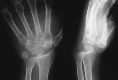
Proximal row carpectomy is a surgical procedure that involves removal of bones of proximal row of the wrist namely Scaphoid, lunate, triquetral and in some cases radial styloid. It is a motion-sparing procedure.

Proximal row carpectomy converts complex wrist joint to a simple hinge joint creating a radiocarpal articulation and preserves about 60-80% of motion & 70-100% grip strength.

However, it has a disadvantage of relative lengthening of extrinsic tendons.

Indications

  • Disabling wrist pain secondary to Scapholunate Advanced Collapse wrist
  • Kienbock’s disease
  • Scaphoid Nonunion
  • Avascular necrosis of scaphoid

Proximal row carpectomy should not be done in severe degenerative changes and rheumatoid arthritis.

Wrist Arthroplasty and wrist fusion are two alternative procedures that must be considered and discussed with the patient.

Xray after Proximal Row Carpectomy
Xray after Proximal Row Carpectomy, Image Credit: Hand Surgery

Technique of Proximal Row Carpectomy

The wrist is opened through a dorsal longitudinal approach through the third compartment. Radioscaphocapitate ligament is preserved so as to prevent ulnar subluxation after the procedure of proximal row carpectomy is performed.

After exposure, traction is applied to the index and long fingers, in a longitudinal direction. This tract facilitates the excision of the proximal carpal row. For bone removal, small rongeurs are used first to excise the middle of each bone. This allows the proximal and distal edges of the proximal carpal bones to collapse together. This makes the rest of the removal of the bones easy along their peripheral edges.

Injury to palmar wrist ligament, triangular fibrocartilage complex, capitate, and pisiform should be avoided. Passive flexion-extension of the hand is done in neutral and slight radial deviation to detect impingement of the trapezium and the radial styloid. Some patients do require radial styloidectomy because of impingement.

The wound is closed and the wrist is immobilized in a well-padded plaster splint for 2 weeks.

Sutures are removed at  2 weeks, the patient is placed in a removable splint, and motion is started.

Related

Spread the Knowledge
14
Shares
 
14
Shares
113   

Filed Under: Procedures

About Dr Arun Pal Singh

Arun Pal Singh is an orthopedic and trauma surgeon, founder and chief editor of this website. He works in Kanwar Bone and Spine Clinic, Dasuya, Hoshiarpur, Punjab.

This website is an effort to educate and support people and medical personnel on orthopedic issues and musculoskeletal health.

You can follow him on Facebook, Linkedin and Twitter

Primary Sidebar

Browse Articles

Deformed pelvis oseomalcia

Osteomalacia Causes and Treatment

Osteomalacia is a  metabolic bone disease where defective mineralization results in bone soften due to a large amount of unmineralized osteoid. Osteomalacia (mollities ossium) is characterized by softening of the bones because of an accumulation of osteoid tissue. Osteoid is the bone matrix which is mineralized to form bone. Like rickets, osteomalacia also affects the […]

Normal Olecranon Bursa and Inflamed Olecranon Bursa or Bursitis

Aspiration of Olecranon Bursa

Aspiration of olecranon bursa is performed to obtain fluid for analysis. A bursa can become inflamed due to infection or for noninfective reasons like trauma, overuse, crystal deposition diseases etc. Non-infective or aseptic causes account for approximately two-thirds of all bursitis diagnoses. Infective or septic bursitis most commonly occurs due to seeding of bacteriae or […]

adhesive capsultis

Adhesive Capsulitis or Frozen Shoulder

Adhesive capsulitis is disease of shoulder that leads to pain and contracture of shoulder. It is commonly referred to as frozen shoulder. Adhesive capsulitis is a poorly understood condition that  is characterized by physical findings like thickening of the synovial capsule and adhesion to surrounding tissues. It is found in 3-5% of the population and […]

apical vertebra and other terms in scoliosis

Idiopathic Scoliosis – Types, Presentation and Treatment

Idiopathic scoliosis is the lateral deviation deformity of the spine where the cause is not known. Because there is also a vertebral rotation and not just deviation, the term rotoscoliosis is also used to describe the deformity. Idiopathic signifies that it is not secondary or due to some deformity and other secondary causes. On plain […]

Representative picture for things to do to achieve fastr fracture healing

7 Tips for Faster Fracture Healing

Fractures are very common in modern life. Reported annual occurrence in the US is about 7 million. Fracture healing can take any time between 4-12 weeks depending on the bone injured and the treatment given. Because of inconvenience and adverse effects on daily activities of living, everyone looks for ways of faster fracture healing. But […]

Knee Joint Aspiration or knee arthrocentesis

Knee Arthrocentesis or Knee Joint Aspiration

Knee arthrocentesis refers to puncture of the knee joint and the aspiration of its synovial fluid. It is used for synovial fluid analysis to aid in diagnosis and also for therapeutic purposes. Knee joint can be approached by multiple approaches [discussed below] Indications of Knee Arthrocentesis Diagnosis Acute monoarticular [that affects a single joint] arthritis […]

Intercondylar View of Knee

Normal Knee X-rays

The knee is an important load-bearing joint of the lower limb. Knee x-rays are done to assess the knee joint pathology. Anteroposterior and lateral views of the knee are most common knee x-rays done. Skyline view is typically used to assess patellofemoral joint and patella-condylar alignment. Knee x-rays in weight-bearing position are taken to study […]

© Copyright: BoneAndSpine.com
Manage Cookie Consent
The site uses cookies. Please accept cookies for a better visiting experience.
Functional Always active
The technical storage or access is strictly necessary for the legitimate purpose of enabling the use of a specific service explicitly requested by the subscriber or user, or for the sole purpose of carrying out the transmission of a communication over an electronic communications network.
Preferences
The technical storage or access is necessary for the legitimate purpose of storing preferences that are not requested by the subscriber or user.
Statistics
The technical storage or access that is used exclusively for statistical purposes. The technical storage or access that is used exclusively for anonymous statistical purposes. Without a subpoena, voluntary compliance on the part of your Internet Service Provider, or additional records from a third party, information stored or retrieved for this purpose alone cannot usually be used to identify you.
Marketing
The technical storage or access is required to create user profiles to send advertising, or to track the user on a website or across several websites for similar marketing purposes.
Manage options Manage services Manage {vendor_count} vendors Read more about these purposes
View preferences
{title} {title} {title}
 

Loading Comments...