• Skip to main content
  • Skip to primary sidebar
  • Skip to footer
  • General Ortho
  • Procedures
  • Spine
  • Upper Limb
  • Lower Limb
  • Pain
  • Trauma
  • Tumors
  • Newsletter/Updates
  • About Us
  • Contact Us

Bone and Spine

Orthopedic health, conditions and treatment

Risser Sign and Its Use in Scoliosis

By Dr Arun Pal Singh

In this article
    • Risser Sign Grading
    • Significance of Risser Sign
    • Drawbacks of Risser Sign

The Risser sign, also called Risser index or Risser classification is a system used to assess and grade skeletal maturity based on the level of ossification and fusion of the iliac crest apophyses.

The apophyses (singular: apophysis) are the normal bony outgrowths which have separate ossification centers and eventually fuse with the bone in time. Iliac crest and greater trochanter are examples of apophyses.

Risser sign is used in scoliosis when corrective surgery is planned.

Scoliotic curves progress with the growth of the skeleton and Risser sign helps to gauge the likelihood of progression of the curve by assessing the growth potential that is present.

As noted previously it is a radiographic evaluation and x-rays are required for evaluating this sign.

Diagram of Risser sign
Risser sign
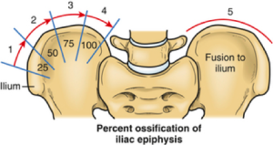
Image Credit: vsrc.com.au

Risser Sign Grading

The sign is gauged on anteroposterior view x-ray of the pelvis.

On x-ray, the amount of ossification of iliac apophysis and is graded from 0 – 5. The process of ossification begins anteriorly and moves to posterior on the apophysis.

The grading is as follows

  • Grade I – Ossification of the lateral 25% of the iliac apophysis
  • Grade II – Ossification of the lateral 50%
  • Grade III- Ossification of the lateral 75% of the apophysis
  • Grade IV – Ossification of the entire [100%] apophysis
  • Grade V – Fusion of ossified epiphysis to the iliac wing

Significance of Risser Sign

As noted before the curves of scoliosis progress during periods of rapid growth.

Thus the progression is more in infancy and adolescence, and they stop growing after spinal maturation is complete.

Males and females stop growing at different ages.

Growth of the spine is completed at approximately 15 years 3 months of age in female adolescents. In male adolescents, the age of this completion is 15 years and 10 months.

This completion or maturation of spinal growth coincides with the completion of ossification in the iliac apophysis.

The ossification of iliac apophysis begins around 15 years in male adolescents and around 14 years in females.

Thus the whole process of ossification from stage  I to stage IV takes around 12-18 months.

After the ossification, the apophysis fuses with iliac crests. The fusion takes around two years in girls and slightly lesser time in boys.

But the progression does not occur after Risser sign grade IV has been achieved.

Some other factors are also responsible for the progression of the curve in addition to skeletal growth. But then also the progression is not as fast as when the skeleton is growing.

For example, if the curve is severe say 50-80° at the time of maturity, it may progress even after the apophysis has fused. This is especially true for the lumbar and thoracic regions.

But the progression occurs far slower than that occurs when growth is pending.

Drawbacks of Risser Sign

Objections have been recently raised to Risser index.

Risser index may not be that reliable. The studies have suggested that ossification starts earlier what we thought previously. Therefore, the predictability and hence reliability is doubtful.

The accuracy of the index is also questioned as the recent data has indicated that this index is not an accurate measure of completed maturity, spinal growth, or progression of the deformity.

In view of these observations, alternate methods of ascertaining the maturity have been suggested.

One such method is ossification of the vertebral ring apophysis.

Before skeletal maturity is attained, the vertebral ring apophysis is absent, or it is incompletely formed.

In some cases, it could be completely formed without fusing to the body.

The vertebral ring apophysis fuses to the body of the vertebra after skeletal maturity.

This fusion occurs by the age of 16 years in female adolescents.

In male adolescents, the age of this fusion is 19 years.

Spread the Knowledge
  • 2
    Shares
  •  
    2
    Shares
  • 2
  •  
  •  
  •  
  •  

Filed Under: Spine

About Dr Arun Pal Singh

Arun Pal Singh is an orthopedic and trauma surgeon, founder and chief editor of this website. He works in Kanwar Bone and Spine Clinic, Dasuya, Hoshiarpur, Punjab.

This website is an effort to educate and support people and medical personnel on orthopedic issues and musculoskeletal health.

You can follow him on Facebook, Linkedin and Twitter

Primary Sidebar

wrist arthrocentesis

Wrist Arthrocentesis – Indications and Procedure

Wrist arthrocentesis is the procedure of aspirating fluid from wrist joint and is used for diagnosis and therapy of various pathologies of the wrist. The wrist joint is anatomically complex and can be approached from any side but the dorsal approach is the preferred site for wrist arthrocentesis. Relevant Anatomy of Wrist Joint Indications of […]

Medial malleolus fracture

A Case of Medial Malleolus Fracture Fixed By Percutaneous Fixation

Medial malleolus fracture is a fracture of the distal medial projection of tibia bone called medial malleolus. Medial malleolus and lateral malleolus contribute forms the socket of the ankle joint where talus articulates. There are three malleoli in the ankle. The medial and the posterior arise from the tibia. A counterpart called lateral malleolus is […]

coracoacrmial arch

Humeral Avulsion of Glenohumeral ligament or HAGL

Humeral avulsion of the glenohumeral ligament or HAGL refers to avulsion of the inferior glenohumeral ligament (IGHL) from its humeral insertion. When it is associated with a bony avulsion fracture, it is called as bony humeral avulsion of the glenohumeral ligament (BHAGL lesion). Humeral avulsion of the glenohumeral ligament is often seen in young men engaged in contact sports […]

What is Fatigue of Material

Fatigue defined as the progressive and localized structural damage that occurs when a material is subjected to cyclic loading. Now if the loads are beyond a certain limit for that material, microscopic cracks will begin to form at the stress concentrators which eventually would reach a critical size, and the material will suddenly fracture. Fatigue […]

Thoracic Spine Anatomy

Thoracic Spine Anatomy

The thoracic spine is also called the dorsal spine and is constituted by the next 12 vertebrae after the cervical spine. These vertebrae are named T1 to T12 (or D1 to D12) from above downwards. If you palpate back of the neck from above downwards in the midline, you would come across a sudden projection […]

Darrach Procedure

Darrach procedure is removal of distal ulna. It was first performed by Darrach in 1911 in New York City. Darrach Procedure is done for- For relief of pain following distal RU disruption and/or RU arthritis; For symptomatic malunion of Colle fracture in elderly patients, especially when stiffness is present Darrach  procedure is generally performed on […]

Spinal Nerves

Spinal Nerves Structure, Types and Function

Spinal nerves are the nerves that originate in the spinal cord and form part of the peripheral nervous system. Spinal nerves are mixed types of nerves. A mixed nerve is one which transmits motor, sensory, and autonomic signals between the spinal cord and the body. In humans, there are 31 spinal nerves on each side […]

Browse Articles

Footer

Pages

  • About
    • Policies
    • Contact Us

Featured Article

Prosthetic Joint Infection

Prosthetic joint infection is a devastating complication of joint replacement surgery and occurs in1–3% of patients. Prosthetic joint infection can … [Read More...] about Prosthetic Joint Infection

Search Articles

© Copyright: BoneAndSpine.com